

Excess soluble vascular endothelial growth factor receptor-1 in amniotic fluid impairs lung growth in rats: linking preeclampsia with bronchopulmonary dysplasia
- PMID: 22003089
- PMCID: PMC3349373
- DOI: 10.1152/ajplung.00294.2011
Excess soluble vascular endothelial growth factor receptor-1 in amniotic fluid impairs lung growth in rats: linking preeclampsia with bronchopulmonary dysplasia
Abstract
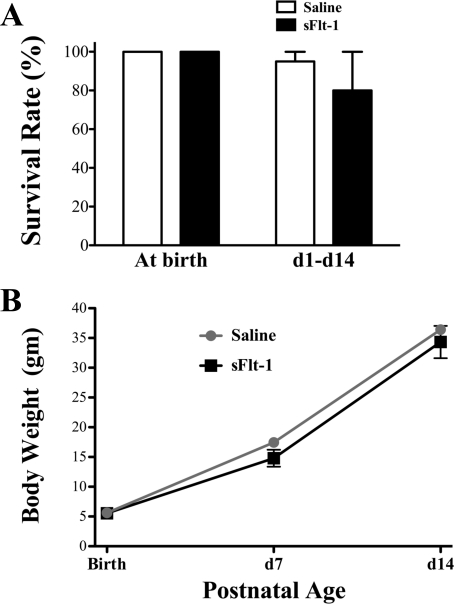
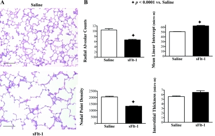
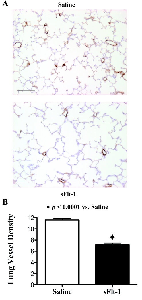
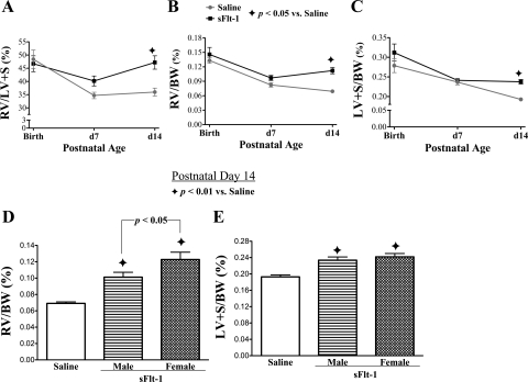
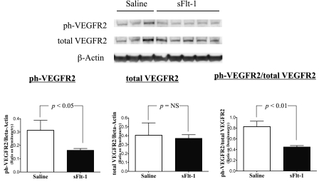
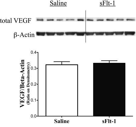
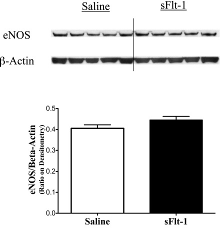
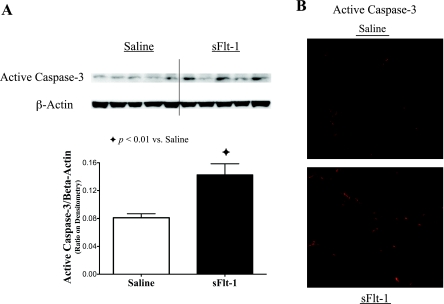
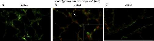
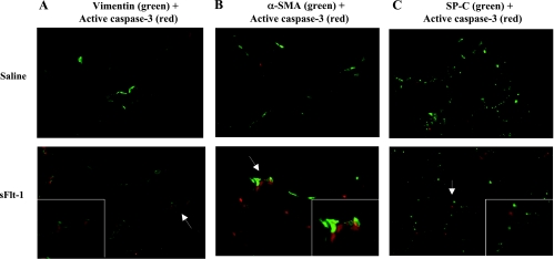
Epidemiological studies have shown that maternal preeclampsia (PE) increases the risk of bronchopulmonary dysplasia (BPD), but the underlying mechanism is unknown. Soluble vascular endothelial growth factor receptor-1 (soluble VEGFR1, known as soluble fms-like tyrosine kinase 1, or sFlt-1), an endogenous antagonist of vascular endothelial growth factor (VEGF), is markedly elevated in amniotic fluid and maternal blood in PE. Therefore, we hypothesized that antenatal exposure to excess sFlt-1 disrupts lung development through impaired VEGF signaling in utero, providing a mechanistic link between PE and BPD. To determine whether increased sFlt-1 in amniotic fluid is sufficient to cause sustained abnormalities of lung structure during infancy, sFlt-1 or saline was injected into amniotic sacs of pregnant Sprague-Dawley rats at 20 days of gestation (term, 22 days). After birth, pups were observed through 14 days of age for study. We found that intra-amniotic sFlt-1 treatment decreased alveolar number, reduced pulmonary vessel density, and caused right and left ventricular hypertrophy in 14-day-old rats. In addition, intra-amniotic sFlt-1 treatment suppressed activation of lung VEGF receptor-2 and increased apoptosis in endothelial and mesenchymal cells in the newborn lung. We conclude that exposure to excess sFlt-1 in amniotic fluid during late gestation causes sustained reductions in alveolarization and pulmonary vascular growth during infancy, accompanied by biventricular hypertrophy suggesting pulmonary and systemic hypertension. We speculate that impaired VEGF signaling in utero due to exposure of high amniotic fluid levels of sFlt-1 in PE disrupts lung growth and contributes to the increased risk of BPD in infants born to mothers with PE.
Figures
References
-
- Akeson AL, Wetzel B, Thompson FY, Brooks SK, Paradis H, Gendron RL, Greenberg JM. Embryonic vasculogenesis by endothelial precursor cells derived from lung mesenchyme. Dev Dyn 217: 11–23, 2000 - PubMed
-
- Anderson PJ, Doyle LW. Neurodevelopmental outcome of bronchopulmonary dysplasia. Semin Perinatol 30: 227–232, 2006 - PubMed
-
- Balasubramaniam V, Maxey AM, Morgan DB, Markham NE, Abman SH. Inhaled NO restores lung structure in eNOS-deficient mice recovering from neonatal hypoxia. Am J Physiol Lung Cell Mol Physiol 291: L119–L127, 2006 - PubMed
-
- Bancalari E, Claure N, Gonzalez A. Patent ductus arteriosus and respiratory outcome in premature infants. Biol Neonate 88: 192–201, 2005 - PubMed
Publication types
MeSH terms
Substances
Grants and funding
LinkOut - more resources
Full Text Sources
Other Literature Sources
Medical
Miscellaneous