. 2011 Nov 17;52(12):8868-77.
doi: 10.1167/iovs.11-8033.

Ectopic proliferation contributes to retinal dysplasia in the juvenile zebrafish patched2 mutant eye

Affiliations

Ectopic proliferation contributes to retinal dysplasia in the juvenile zebrafish patched2 mutant eye

Jonathan Bibliowicz et al. Invest Ophthalmol Vis Sci. .

Abstract

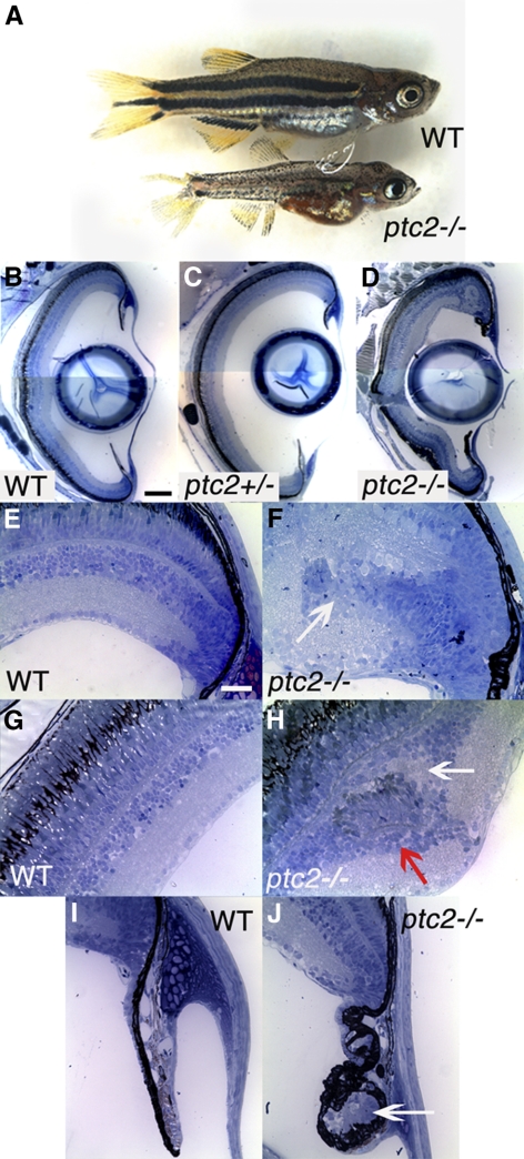
Purpose: Patched is a well-studied tumor suppressor and negative regulator of the Hedgehog (Hh) pathway. Earlier work in this laboratory has shown that embryonic zebrafish patched2 (ptc2) mutant retinas possess an expanded ciliary marginal zone (CMZ) and phenotypes similar to those in human patients with basal cell naevus syndrome (BCNS), a congenital disorder linked to mutations in the human PTCH gene. This study extends the analysis of retinal structure and homeostasis in ptc2-/- mutants to juvenile stages, to determine whether Patched 2 function is essential in the postembryonic eye.

Methods: Histologic, immunohistochemical, and molecular analyses were used to characterize retinal defects in the 6-week-old juvenile ptc2-/- retina.

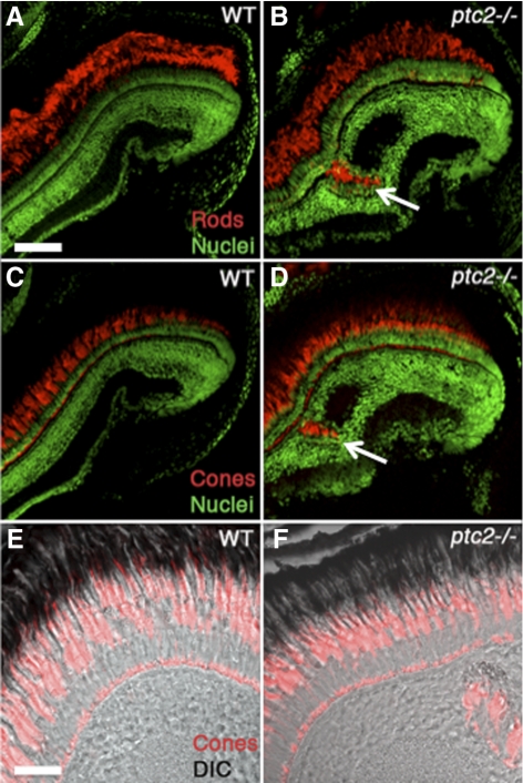
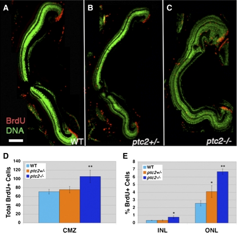
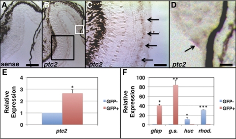
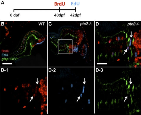
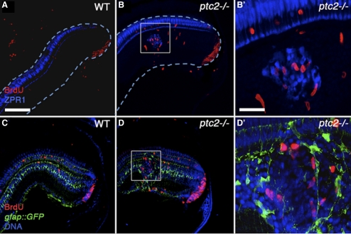
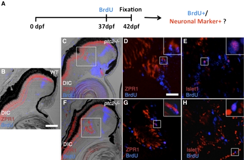
Results: Juvenile ptc2-/- mutants exhibited peripheral retinal dysplasias that included the presence of ectopic neuronal clusters in the inner nuclear layer (INL) and regions of disrupted retinal lamination. Retinal dysplasias were locally associated with ectopic proliferation. BrdU/EdU labeling and immunohistochemistry assays demonstrated that a population of ectopically proliferating cells gave rise to the ectopic neuronal clusters in the INL of ptc2-/- mutants and that this contributed to retinal dysplasia in the mutant eye.

Conclusions: These results demonstrate a direct link between overproliferation and retinal dysplasia in the ptc2-/- juvenile retina and establish ectopic proliferation as the likely cellular underpinning of retinal dysplasia in juvenile ptc2-/- mutants.


Figures

Figure 1.
Figure 1.
Juvenile ptc2−/− mutants possess peripheral retinal dysplasias and abnormalities at the ciliary zone. A small percentage of ptc2−/− mutants (∼2%) reach 6 weeks of age. ptc2−/− mutants are smaller than their wild-type siblings and have a disrupted pigmentation pattern (A). Histologic analyses of wild-type (B, E, G, I), ptc2+/− (C), and ptc2−/− (D, F, H, J) 6-week retinas. Retinal organization appeared normal in ptc2+/− mutants (C). In ptc2−/− mutants, retinal disorganization was found in the dorsal peripheral retina, whereas the rest of the retina was laminated normally (D). Within the dorsal peripheral retina of ptc2−/− mutants, ectopic neurons that appeared to be continuous with the CMZ and disrupted lamination were detected (F, arrow; n = 4/10). Ectopic neuronal clusters in the INL were also observed (H, n = 3/10). Neuronal clusters contained photoreceptor outer segments (H, red arrow) and were associated with an ectopic plexiform layer (H, white arrow). Morphologic abnormalities and ectopic cells (J, arrow) were observed in the ciliary zone (n = 3/10). Scale bar: (BD) 100 μm; (EJ) 25 μm.
Figure 2.
Figure 2.
Ectopic neuronal clusters in juvenile ptc2−/− retinas contain rod and cone photoreceptors. Both ZPR3-expressing rods (B, arrow) and ZPR1-expressing red/green double cones (D, arrow) were detected in the ectopic neuronal clusters in ptc2−/− retinas, whereas in wild-type retinas (A, C), photoreceptors were restricted to the ONL. Higher magnification views of cone photoreceptor morphology in wild-type (E) and ptc2−/− (F) retinas. Scale bar: (AD) 100 μm; (E, F) 50 μm.
Figure 3.
Figure 3.
The ptc2−/− retina is overproliferative. BrdU incorporation (2 hours) at 6 weeks revealed an increase in the number of proliferative cells in the ptc2−/− retina (C) when compared to wild-type (A) (n = 4). The total number of BrdU+ cells in the CMZ was increased by 54% in ptc2−/− mutants when compared with wild-type siblings (**P < 0.001, n = 4) (D). The percentage of BrdU+ cells in the INL (2.30-fold, *P < 0.01, n = 4) and ONL (2.62-fold, **P < 0.001, n = 4) were significantly increased in ptc2−/− mutants when compared to wild-type siblings (E). A statistically significant increase in the percentage of BrdU+ cells was also observed in the ONL of ptc2+/− mutants (B, E) (1.54-fold, *P < 0.01, n = 4) when compared to wild-type (A, E). Scale bar: 100 μm.
Figure 4.
Figure 4.
ptc2 is expressed in progenitor/stem cell populations of the juvenile zebrafish retina. In situ hybridization for the ptc2 transcript in 6-week-old juvenile wild-type retinas. Staining was absent throughout the retina when hybridization was performed using a ptc2 sense control probe (A). (B) ptc2 expression was detected in a population of cells in the INL (C; High magnification of the black box) and in the retinal margin (D; high magnification of white box), where CMZ progenitors reside. qRT-PCR analysis of ptc2 expression from FACS-sorted GFP+ Müller glia cells isolated from a gfap:GFP transgenic line revealed enrichment of the ptc2 transcript in GFP+ Müller glia when compared to GFP, non-Müller glial cells (E, *P < 0.01, n = 3). GFP+ cells were enriched for the Müller glial markers gfap (***P < 0.01, n = 3) and glutamine synthetase (**P < 0.001, n = 3), and GFP cells were enriched for the neuronal markers huc (*P < 0.01, n = 3) and rhodopsin (***P < 0.0001, n = 3) (F). Scale bars: (A, B) 100 μm; (C) 50 μm; (D) 25 μm.
Figure 5.
Figure 5.
Continually proliferative cells in regions of retinal dysplasia in ptc2−/− mutants. A BrdU/EdU double-labeling experiment was performed on wild-type gfap:GFP (B) and ptc2−/−;gfap:GFP mutant retinas (C, D, D13). The fish were exposed to BrdU for 8 hours at 40 dpf, returned to their tanks for 2 days, injected with EdU at 42 dpf (6 weeks), and fixed for BrdU and EdU immunohistochemistry and detection (A). In wild-type retinas, cells that were proliferative at the time of fixation (EdU+) were mostly confined to the CMZ, whereas cells that were proliferative 2 days before fixation (BrdU+) had incorporated into the retina. In addition, a few GFP+ Müller glia were also either BrdU+ or EdU+ (B). In ptc2−/−;gfap:GFP retinas, in addition to BrdU+/GFP+ or EdU+/GFP+ Müller glia, BrdU+/EdU+ double-labeled cells were detected in the peripheral retina, and these did not express GFP (C, and white arrows in high magnification of boxed region in D and in D13, n = 3/3). Scale bars: (B, C) 100 μm; (D, D13) 20 μm.
Figure 6.
Figure 6.
Ectopic neuronal clusters in regions of retinal dysplasia are locally associated with ectopic proliferation. Immunohistochemical analysis of wild-type retinas after a 2-hour exposure to BrdU revealed few proliferative cells in the INL (A, C), whereas in dysplastic regions of the ptc2−/− retina, BrdU+ cells were locally associated with ectopic ZPR1-expressing red/green double cones in the INL (B). (B′: high magnification of white box.) Analysis of adjacent sections from the same individuals showed that these proliferative cells were not Müller glia, as they did not label with GFP in the gfap:GFP transgenic background (D). (D′: high magnification of white box). Scale bar: 100 μm.
Figure 7.
Figure 7.
New neurons are generated by ectopic proliferation in the dysplastic ptc2−/− juvenile retina. A combined BrdU incorporation and immunohistochemical assay to determine whether ectopically proliferative cells generate new neurons in dysplastic regions of the ptc2−/− retina (A). Fish were exposed to BrdU for 8 hours at 37 dpf, returned to their tanks for 5 days, and fixed for immunohistochemistry at 42 dpf (6 weeks). In wild-type siblings (B), BrdU+ cells were detected in a narrow band within the differentiated retina, corresponding to CMZ-derived cells that were proliferative 5 days before fixation, as well as in scattered rod progenitors located in the ONL. Red/green double cones, labeled by ZPR1, were restricted to the ONL. In two different ptc2−/− retinas, BrdU+ cells colocalized with ZPR1 (C, F, high magnification of boxed areas in D, G). In adjacent sections from the same individuals, BrdU+ cells were also positive for Islet1, a marker of retinal ganglion cells, and subsets of differentiated amacrine, bipolar, and horizontal cells (E, H). (D, E, G, H, insets) Higher magnification of the respective boxed regions in each panel, showing co-localization of BrdU and the neuronal marker. Scale bars: (B, C, F) 100 μm; (D, E, G, H) 20 μm.

Similar articles

Cited by

References

    1. Ekker SC, Ungar AR, Greenstein P, et al. Patterning activities of vertebrate hedgehog proteins in the developing eye and brain. Curr Biol. 1995;5:944–955 - PubMed
    1. Ericson J, Muhr J, Placzek M, Lints T, Jessell TM, Edlund T. Sonic hedgehog induces the differentiation of ventral forebrain neurons: a common signal for ventral patterning within the neural tube. Cell. 1995;81:747–756 - PubMed
    1. Dahmane N, Ruiz i Altaba A. Sonic hedgehog regulates the growth and patterning of the cerebellum. Development. 1999;126:3089–3100 - PubMed
    1. Jiang J, Hui CC. Hedgehog signaling in development and cancer. Dev Cell. 2008;15:801–812 - PMC - PubMed
    1. Ohnuma S, Hopper S, Wang KC, Philpott A, Harris WA. Co-ordinating retinal histogenesis: early cell cycle exit enhances early cell fate determination in the Xenopus retina. Development. 2002;129:2435–2446 - PubMed

Publication types

MeSH terms

LinkOut - more resources