. 2011 Nov 15;187(10):5062-8.
doi: 10.4049/jimmunol.1102001. Epub 2011 Oct 14.

MicroRNA-125b potentiates macrophage activation

Affiliations

MicroRNA-125b potentiates macrophage activation

Aadel A Chaudhuri et al. J Immunol. .

Abstract

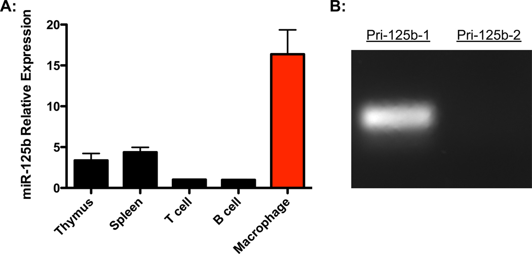
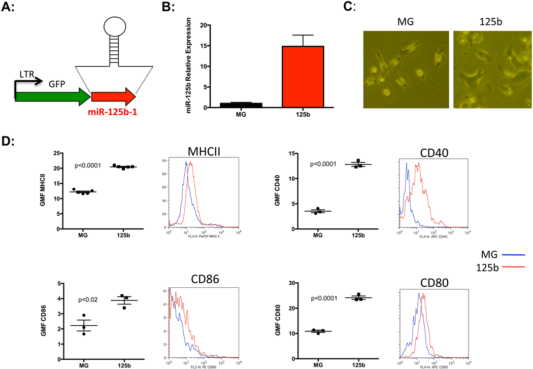
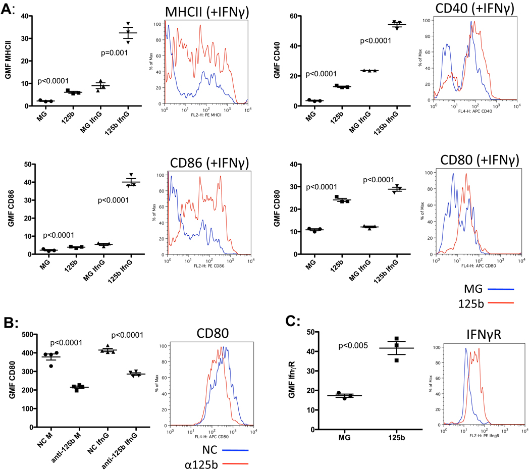
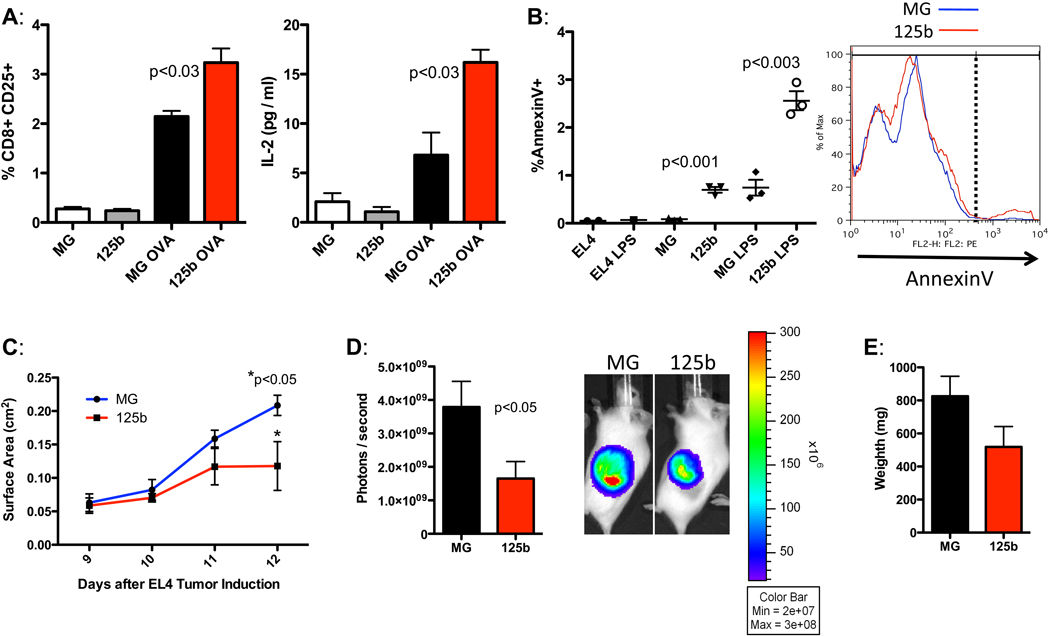
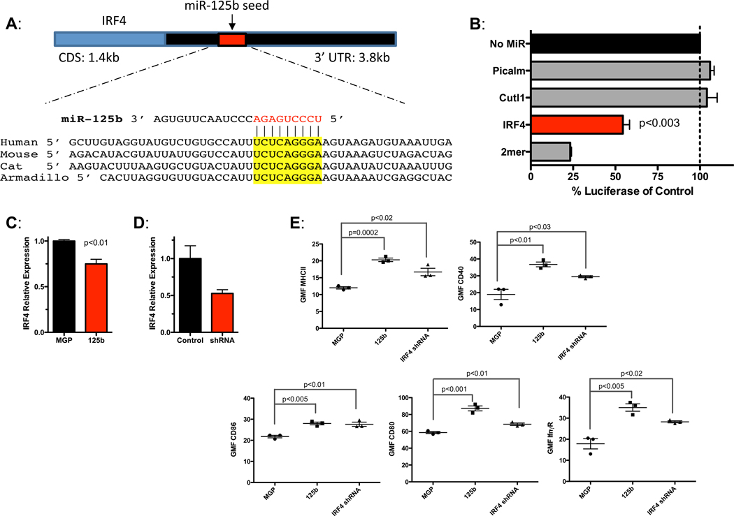
MicroRNA (miR)-125b expression is modulated in macrophages in response to stimulatory cues. In this study, we report a functional role of miR-125b in macrophages. We found that miR-125b is enriched in macrophages compared with lymphoid cells and whole immune tissues. Enforced expression of miR-125b drives macrophages to adapt an activated morphology that is accompanied by increased costimulatory factor expression and elevated responsiveness to IFN-γ, whereas anti-miR-125b treatment decreases CD80 surface expression. To determine whether these alterations in cell signaling, gene expression, and morphology have functional consequences, we examined the ability of macrophages with enhanced miR-125b expression to present Ags and found that they better stimulate T cell activation than control macrophages. Further indicating increased function, these macrophages were more effective at killing EL4 tumor cells in vitro and in vivo. Moreover, miR-125b repressed IFN regulatory factor 4 (IRF4), and IRF4 knockdown in macrophages mimicked the miR-125b overexpression phenotype. In summary, our evidence suggests that miR-125b is at least partly responsible for generating the activated nature of macrophages, at least partially by reducing IRF4 levels, and potentiates the functional role of macrophages in inducing immune responses.


Figures

Figure 1
Figure 1
MiR-125b expression is enriched in macrophages. A) Relative expression of miR-125b in immune tissues and cells assessed by quantitative PCR. Data represents the mean with SEM of 3 biological replicates per group. B) Expression of the miR-125b primary transcripts, pri-125b-1 and pri-125b-2, in bone marrow derived macrophages (BMMs). Data is representative of two independent experiments.
Figure 2
Figure 2
MiR-125b enhances basal macrophage activation. A) Retroviral vector design for over-expression of miR-125b-1. B) Relative expression of miR-125b in BMMs after transduction with MG or MG-miR-125b expressing vector. C) Morphology of control MG or miR-125b over-expressing BMMs. Data represent five independent experiments. D) Geometric mean fluorescence (GMF) of MHCII, CD40, CD86 and CD80 are shown. Representative plots obtained from flow cytrometric analyses are also shown for each marker. Data is the mean with SEM of 3–5 samples per group and is representative of two independent experiments.
Figure 3
Figure 3
MiR-125b increases macrophage response to IFNγ. A) Surface expression of MHCII, CD40, CD86, and CD80 in response to media alone or IFNγ. A representative flow cytometric plot of the IFNγ treated samples is shown for each factor. B) Raw264.7 macrophages electroporated with control (NC) or anti-miR-125b were subjected to flow cytometry for the surface expression of CD80. A representative FACS plot of the media-treated samples shown. C) Surface expression of IFNγR in control (MG) versus miR-125b over-expressing macrophages. A representative FACS plot is shown. All data shown represents the mean expressed with SEM of three samples per group and is representative of two independent experiments.
Figure 4
Figure 4
MiR-125b enhances macrophage function. A) BMMs expressing the vectors MG or MG-125b were co-cultured with ovalbumin-specific OT1 T cells with or without ovalbumin for 72 hours. The percent CD8+CD25+ T cells are shown in the left panel. Concentration of IL-2 (pg/ml) produced by the T cells in the supernatant is shown in the right panel. Data represents the mean with SEM of 3 biological replicates per group. B) The percent AnnexinV+ EL4-Fluc cells after 94 hours of co-culture with control or miR-125 over-expressing macrophages in the presence of media alone or lipopolysaccharide (LPS). A representative flow cytometric plot of the LPS-treated group is shown. Data is expressed in mean with SEM of 1–3 experimental samples per group. C–E) EL4-Fluc cells were subcutaneously co-injected with LPS-activated control or miR-125b over-expressing macrophages into albino C57Bl/6 mice. Tumor surface area in cm2 was monitored from day 9–12 (C). The relative intensity of luminescence (D) and weight (E) of the EL4 tumors were measured on day 12. Data represents the mean plotted with SEM of eight mice per group. Representative of two independent experiments.
Figure 5
Figure 5
IRF4 is a target of miR-125b in macrophages. A) IRF4 contains a conserved miR-125b target site. B) Luciferase reporters carrying the 3’UTR of IRF4, Picalm (negative control), Cutl1 (negative control) or 2mer (positive control) were co-transfected into 293T cells with β-gal reporter and +/− miR-125b. The relative luciferase activity of each reporter in the presence of miR-125b is shown relative to the no microRNA control. C) RAW264.7 macrophages were transduced with either a control (MGP) or miR-125b expressing vector, or with D) control (NC1) or IRF4 shRNA expressing vector. RNA was harvested and L32-normalized IRF4 levels were determined by qPCR. E) BMMs expressing MGP, MGP-125b, or shRNA against IRF4 were measured for surface expression of the activation markers MHCII, CD40, CD86, CD80 and IFNγR. Geometric Mean Fluorescence (GMF) measured by flow cytometry is shown. All data represents the mean with SEM of 3 samples per group and is representative of two independent experiments.

References

    1. Murphy KP, Travers P, Walport M, Janeway C. Janeway's immunobiology. New York: Garland Science; 2008.
    1. Kindt TJ, Goldsby RA, Osborne BA, Kuby J. Kuby immunology. New York: W.H. Freeman; 2007.
    1. O'Connell RM, Rao DS, Chaudhuri AA, Baltimore D. Physiological and pathological roles for microRNAs in the immune system. Nat Rev Immunol. 2010;10:111–122. - PubMed
    1. O'Neill LA, Sheedy FJ, McCoy CE. MicroRNAs: the fine-tuners of Toll-like receptor signalling. Nat Rev Immunol. 2011;11:163–175. - PubMed
    1. Androulidaki A, Iliopoulos D, Arranz A, Doxaki C, Schworer S, Zacharioudaki V, Margioris AN, Tsichlis PN, Tsatsanis C. The kinase Akt1 controls macrophage response to lipopolysaccharide by regulating microRNAs. Immunity. 2009;31:220–231. - PMC - PubMed

Publication types

MeSH terms