Review
. 2011 Nov 8;50(44):9424-33.
doi: 10.1021/bi201157t. Epub 2011 Oct 17.

Two distinct catalytic strategies in the hepatitis δ virus ribozyme cleavage reaction

Affiliations
Review

Two distinct catalytic strategies in the hepatitis δ virus ribozyme cleavage reaction

Barbara L Golden. Biochemistry. .

Abstract

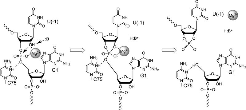
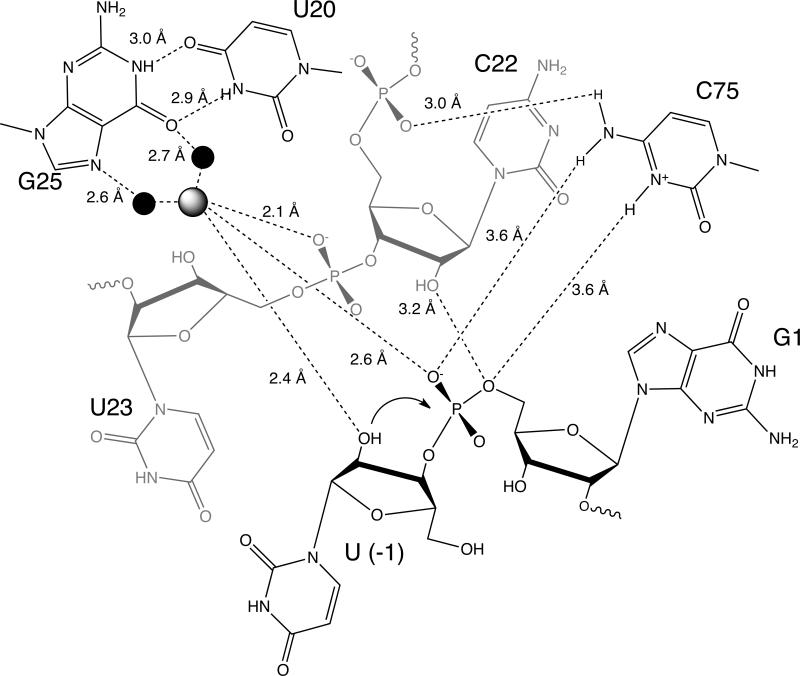
The hepatitis delta virus (HDV) ribozyme and related RNAs are widely dispersed in nature. This RNA is a small nucleolytic ribozyme that self-cleaves to generate products with a 2',3'-cyclic phosphate and a free 5'-hydroxyl. Although small ribozymes are dependent on divalent metal ions under biologically relevant buffer conditions, they function in the absence of divalent metal ions at high ionic strengths. This characteristic suggests that a functional group within the covalent structure of small ribozymes is facilitating catalysis. Structural and mechanistic analyses have demonstrated that the HDV ribozyme active site contains a cytosine with a perturbed pK(a) that serves as a general acid to protonate the leaving group. The reaction of the HDV ribozyme in monovalent cations alone never approaches the velocity of the Mg(2+)-dependent reaction, and there is significant biochemical evidence that a Mg(2+) ion participates directly in catalysis. A recent crystal structure of the HDV ribozyme revealed that there is a metal binding pocket in the HDV ribozyme active site. Modeling of the cleavage site into the structure suggested that this metal ion can interact directly with the scissile phosphate and the nucleophile. In this manner, the Mg(2+) ion can serve as a Lewis acid, facilitating deprotonation of the nucleophile and stabilizing the conformation of the cleavage site for in-line attack of the nucleophile at the scissile phosphate. This catalytic strategy had previously been observed only in much larger ribozymes. Thus, in contrast to most large and small ribozymes, the HDV ribozyme uses two distinct catalytic strategies in its cleavage reaction.


Figures

Figure 1
Figure 1. The structure of the HDV ribozyme
A. Secondary structure of the HDV ribozyme. The HDV ribozyme folds into a pseudoknot containing five core paired regions (P1-P4, P1.1). In the trans-acting version of the ribozyme, a short substrate, U(-1) to A8 here, base pairs to the ribozyme to form the P1 helix. The cleavage site is indicated by an asterisk. The junctions J1.1/3 and J4/2 contribute key nucleobases to the active site of the ribozyme. B. Tertiary structure of the HDV ribozyme (3NKB). The active site is located at the junction of helices P1, P1.1 and P3. Key residues are labeled. A largely hydrated active site Mg2+ ion is shown as spheres. Figure adapted from ().
Figure 2
Figure 2. The catalytic mechanism of the HDV ribozyme
In the substrate bound state (left), C75 is protonated and donates a hydrogen bond to the 5’-O of G1. The active site metal ion is directly coordinated to the pro-RP oxygen of G1 and the 2’-hydroxyl of U(-1). Metal binding to the nucleophile facilitates deprotonation by a yet unknown base (:B). C75+ and the active site Mg2+ ion have the potential to stabilize a phosphorane intermediate (middle) generated by attack of the 2’-O on the scissile phosphate. This intermediate is resolved to form the 2’,3’-cyclic phosphate on U(-1) and a free 5’-hydroxyl on G1 (right).
Figure 3
Figure 3. The cleavage site of the hammerhead ribozyme docked into the active site of the HDV ribozyme
The HDV ribozyme active site is shown in green and the cleavage site is shown in pink. A. Key atoms in the cleavage site, including the 2’-hydroxyl of U(-1), the pro-R P oxygen of G1, and the 5’-hydroxyl of G1, are held in place by at least two hydrogen-bonds, or metal mediated interactions. G25 is positioned directly under G27, and it is not labeled. B. There is extensive shape complementarity between the cleavage site and the active site. Molecular surfaces for the cleavage site (pink) and the active site (green) were generated independently. The active site metal ion and its hydration shell are shown as spheres.
Figure 4
Figure 4. The active site of the product-bound ribozyme is similar to the active site of the inhibitor-bound ribozyme
A. The active site of the product bound ribozyme (yellow), 1CXO, was superposed on the active site of the inhibitor-bound ribozyme (green), 3NKB. Only minor differences were observed between the two structures. The structure of the inhibited ribozyme is consistent with the electron density of the product-bound ribozyme. Thus the observed differences are likely due to the weak electron density in this region of the 1CXO structure. B. The active site of the C75U mutant (red), 1SJ3, is significantly different than the active site of the inhibitor-bound ribozyme (green), 3NKB. Mutation of C75 to a U disrupts a critical hydrogen bond network that organizes the active site. The active site of the C75U mutant is therefore significantly less compact. Figure adapted from ().
Figure 5
Figure 5. Hydrogen bonds and metal mediated interactions in the HDV ribozyme active site
The cleavage site dinucleotide from the hammerhead ribozyme was docked into the active site of the HDV ribozyme as previously described. Distances for hydrogen bonds and metal mediated interactions in the active site are shown. Figure adapted from ().
Figure 6
Figure 6. Comparison of metal binding by a standard G•U and a reverse G•U wobble
Standard G•U wobble base pairs (left) are often associated with major groove Mg(H2O)62+ binding sites (PDBID 1HR2). G25 and U20 base pair in the reverse G•U wobble geometry (right). This helps to create a binding site for a largely hydrated Mg2+ ion in the minor groove. The minor groove is on the left in both panels.
Figure 7
Figure 7. The active site metal binding pocket can accommodate a variety of cations
The active site Mg2+ (yellow, marked with an asterisk) observed in the inhibitor-bound ribozyme (3NKB) overlaps with the binding sites for Mg2+ (yellow), Mn2+ (violet), Ba2+ (green), Tl+ (red) and Co(NH3) 3+6 (blue and pink) observed in the structures of the C75U mutant (1SJ3, 1SJF, 2OIH, 1VBZ, 1VBY).

References

    1. Fedor MJ. Comparative enzymology and structural biology of RNA self-cleavage. Ann. Rev. Biophys. 2009;38:271–299. - PubMed
    1. Reiter NJ, Chan CW, Mondragon A. Emerging structural themes in large RNA molecules. Curr. Opin. Struct. Biol. 2011;21:319–326. - PMC - PubMed
    1. Liberman JA, Wedekind JE. Base ionization and ligand binding: how small ribozymes and riboswitches gain a foothold in a protein world. Curr. Opin. Struct. Biol. 2011;21:327–334. - PMC - PubMed
    1. DeRose VJ. Metal ion binding to catalytic RNA molecules. Curr. Opin. Struct. Biol. 2003;13:317–324. - PubMed
    1. Isom DG, Cannon BR, Castaneda CA, Robinson A, Garcia-Moreno B. High tolerance for ionizable residues in the hydrophobic interior of proteins. Proc. Natl. Acad. Sci. U. S. A. 2008;105:17784–17788. - PMC - PubMed

Publication types