. 2011 Oct 19;101(8):1816-24.
doi: 10.1016/j.bpj.2011.08.034.

Positive feedback loops for factor V and factor VII activation supply sensitivity to local surface tissue factor density during blood coagulation

Affiliations

Positive feedback loops for factor V and factor VII activation supply sensitivity to local surface tissue factor density during blood coagulation

A N Balandina et al. Biophys J. .

Abstract

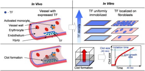
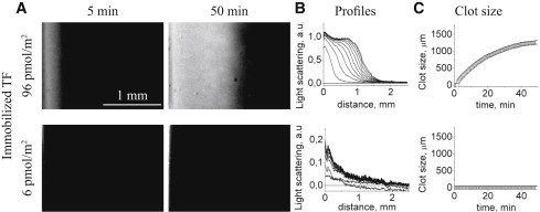
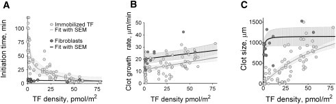
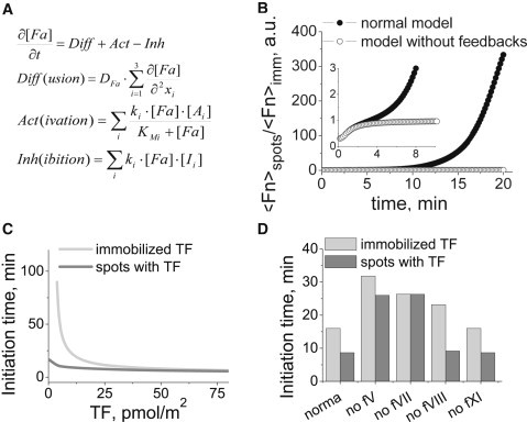
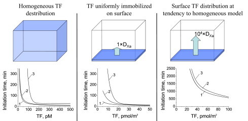
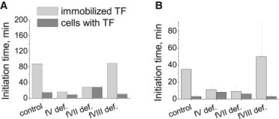
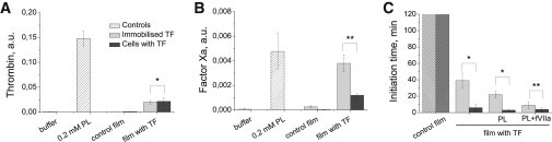
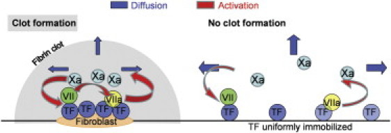
Blood coagulation is triggered not only by surface tissue factor (TF) density but also by surface TF distribution. We investigated recognition of surface TF distribution patterns during blood coagulation and identified the underlying molecular mechanisms. For these investigations, we employed 1), an in vitro reaction-diffusion experimental model of coagulation; and 2), numerical simulations using a mathematical model of coagulation in a three-dimensional space. When TF was uniformly immobilized over the activating surface, the clotting initiation time in normal plasma increased from 4 min to >120 min, with a decrease in TF density from 100 to 0.7 pmol/m(2). In contrast, surface-immobilized fibroblasts initiated clotting within 3-7 min, independently of fibroblast quantity and despite a change in average surface TF density from 0.5 to 130 pmol/m(2). Experiments using factor V-, VII-, and VIII-deficient plasma and computer simulations demonstrated that different responses to these two TF distributions are caused by two positive feedback loops in the blood coagulation network: activation of the TF-VII complex by factor Xa, and activation of factor V by thrombin. This finding suggests a new role for these reactions: to supply sensitivity to local TF density during blood coagulation.


Figures

Figure 1
Figure 1
Blood clotting activation on TF-expressing surfaces in a vessel (in vivo) and in a reaction-diffusion experimental model (in vitro). Under normal conditions, TF is expressed by subendothelial cells only. In many pathological conditions, such as inflammation and cancer, TF can also appear on the surface of blood cells and endothelial cells, either isolated or in monolayers. TF density varies over a wide range. By analogy, coagulation in the in vitro model system used herein was activated by films with either uniformly distributed TF or TF-bearing cells. In both cases, the average surface TF density varied over two orders of magnitude. Spatial clot growth was recorded by means of light scattering. Initiation times, clot growth rates, and clot sizes after 40 min were determined as shown.
Figure 2
Figure 2
Effect of uniformly immobilized TF on fibrin clot growth in normal plasma depends on the TF density. Typical experimental results for platelet-free plasma coagulation activated by TF at 96 and 6 pmol/m2 are shown. (A) Images of fibrin clots for different TF densities as registered by light scattering are shown. Clotting was performed in thin layers of plasma from healthy donors. The activating surface was located on the vertical wall of the experimental chamber and is shown as a vertical stripe at the left end of each image. During the experiment (2 h), large fibrin clots were formed on the films with high TF densities, but no clot formation was detected with the lowest TF density. For lower TF densities, clot formation was delayed and clot size was smaller. (B) Corresponding profiles of clot growth (note the different scales). Each curve (profile) corresponds to a different time point during clot growth; the left and lowest curve corresponds to 5 min after recalcification, and the subsequent curves are separated by 5-min intervals. As the clot grows and increases in size, the curves shift upward (implying that the clot becomes denser) and to the right (the clot becomes larger). The first 50 min are shown. (C) Clot size as a function of time. The plots were obtained using the series of profiles shown in panel B.
Figure 3
Figure 3
Clotting activation by fibroblasts was independent of their density, whereas clot formation by uniformly immobilized activators was sensitive to TF density. Initiation time (A), clot growth rate (B), and clot size after 40 min (C) are plotted as functions of average TF density for uniformly distributed TF (solid gray circles) and TF-expressing cells (solid dark gray circles). Normal plasma from 23 donors was used. For data approximation, the mathematical functions used were a second-order exponential (A), hyperbolic (B), and hyperbolic plus linear (C). Approximation curves are supplemented with the corresponding mean ± SE.
Figure 4
Figure 4
fV activation by thrombin and fVII activation by fXa are essential for clotting system recognition of the local TF concentration. A detailed mechanism-driven mathematical model was used. (A) General equations (F, factor; A, activator; I, inhibitor; t, time; x, spatial coordinate). (B) The ratio of total fibrin generated upon clotting activation by spotted TF to that generated upon stimulation with the same quantity of uniformly distributed TF in the models with and without feedback loops. (C) Dependence of the initiation time on the average surface TF density. Clotting was activated by uniformly distributed TF (gray lines) or by rectangular spots (dark gray lines) with a high TF density (60 × 15 μm size and 500 pmol/m2 TF density) in a 3D region. To evaluate the role of feedback loops in clotting sensitivity to TF distribution, we individually switched off positive feedback loops for fV, fVII, fVIII, and fXI activation by setting the rates of these reactions to zero in our computer simulations (D). The average TF density for this series of simulations was 12.5 pmol/m2.
Figure 5
Figure 5
Spatial TF distribution defines the role of positive feedback for fVII activation in clotting. To compare the roles of fV and fVII feedback loops, initiation time dependencies on TF concentration and density were calculated for normal plasma and plasma without either fVII or fV activation. A detailed, mechanism-driven mathematical model was used. (Left) When TF was distributed homogeneously, only the fV activation feedback loop was essential for clotting. (Middle) When immobilized TF was uniformly distributed over the surface in 3D computer simulations, the feedback loops for both fV and fVII activation influenced the initiation time. (Right) TF was uniformly immobilized at the surface, and the fXa diffusion coefficient was increased by 104 to mimic homogeneous TF distribution. The role of positive feedback in the fVII activation of clotting disappeared, suggesting that fVII activation by fXa is important for the clotting process only when fXa is removed by diffusion; thus, additional fVIIa activated fXa and compensated for its diffusion.
Figure 6
Figure 6
Positive feedback loops for fV and fVII activation render clotting sensitive to surface TF distribution. To validate the mathematical model prediction that the clotting system can recognize local TF concentration only after fV and fVII activation, we performed experiments with fV-, fVII-, and fVIII-deficient plasma. The mathematical model (A) and experimental data (B) are shown. The average TF density was 6.25 pmol/m2 for the mathematical model and 8 pmol/m2 for the experiments. Plots show the initiation times for the control plasma and the fV-, fVII-, and fVIII-deficient plasmas. Immobilized TF and TF-bearing cells were used to activate clotting. For the control plasma, a mixture of fV- and fVII-deficient plasma at a 1:1 volume ratio was used. To enhance clot activation, fV-deficient plasma (<1%) was supplemented with fVa (0.1% of the normal experimental fV concentration and 1% for the model), and fVII-deficient plasma was supplemented with <1% VIIa (1% of the normal fVII concentration). Control and fVIII-deficient plasmas were sensitive to TF distribution in the clotting process, whereas fV- and fVII-deficient plasma showed only small differences between activation by uniform immobilized TFs and fibroblasts. Experimental data are shown with the corresponding SD (n = 3÷5).
Figure 7
Figure 7
Effect of plasma and cell surface lipids on the sensitivity to local TF density during coagulation. (A) Thrombin production by prothrombinase assembled on PL, films with fibroblasts, or films with immobilized TFs. Shown are the mean ± SD, n = 8÷16. (B) fXa production by intrinsic tenase assembled on either PL, films with fibroblasts, or films with immobilized TFs. Shown are the means ± SD, n = 4÷6. (C) Effect of PL (0 and 10 μM) and fVIIa (0 and 10 nM) on the clotting initiation time. Clot formation was initiated by films with fibroblasts or immobilized TFs, both with an average surface density of 8 pmol/m2. Control films are films without TFs. Shown are the means ± SD, n = 3÷7. Asterisks show a significant difference, and double asterisks show a lack thereof, as calculated using an unpaired, two-sample t-test at p = 0.05.
Figure 8
Figure 8
Positive feedback loops supply sensitivity to surface TF distribution during coagulation. (Left) When clotting is activated by cells with a high surface TF density, fX activation is more important than the removal of fXa due to diffusion and inhibition. This result is reinforced by the feedback loop for fVII activation, and the clot rapidly forms near the cells and propagates from them. (Right) In contrast, if the same quantity of TF is distributed uniformly across the surface, then fXa removal and inactivation predominate, and clot formation is prevented.

Similar articles

Cited by

References

    1. Monroe D.M., Key N.S. The tissue factor-factor VIIa complex: procoagulant activity, regulation, and multitasking. J. Thromb. Haemost. 2007;5:1097–1105. - PubMed
    1. Butenas S., Mann K.G. Kinetics of human factor VII activation. Biochemistry. 1996;35:1904–1910. - PubMed
    1. Hemker H.C., Al Dieri R., Béguin S. Thrombin generation, a function test of the haemostatic-thrombotic system. Thromb. Haemost. 2006;96:553–561. - PubMed
    1. Rand M.D., Lock J.B., Mann K.G. Blood clotting in minimally altered whole blood. Blood. 1996;88:3432–3445. - PubMed
    1. Monroe D.M., Hoffman M., Roberts H.R. Transmission of a procoagulant signal from tissue factor-bearing cell to platelets. Blood Coagul. Fibrinolysis. 1996;7:459–464. - PubMed

Publication types