. 2011 Nov;27(11):477-84.
doi: 10.1016/j.kjms.2011.06.010. Epub 2011 Aug 25.

Glutathione-S-transferase enhances proliferation-migration and protects against shikonin-induced cell death in breast cancer cells

Affiliations

Glutathione-S-transferase enhances proliferation-migration and protects against shikonin-induced cell death in breast cancer cells

Shiping He et al. Kaohsiung J Med Sci. 2011 Nov.

Abstract

Glutathione-S-transferase (GST) is a cytoplasmic protein responsible for detoxification, but the effect of the enzyme on cell biological events, including proliferation and migration, has never been reported. Thus, we evaluated the detoxification effect of in vitro-applied GST on cancer cell proliferation and migration. Assays for proliferation and migration of human breast cancer cells in the presence of GST were carried out. Binding of GST on the surface of the cancer cells was studied by flow cytometry. Detoxification through GST pathway was studied in the presence of shikonin. The effective dosage of GST in enhancement of cell proliferation was 10-50 nM, and the cell migration could be significantly enhanced after 6 hours in the presence of 2-50 nM GST. Therefore, overall cell proliferation and migration could be enhanced in the presence of 10nM or greater concentration of GST, and 15 μM shikonin-induced toxification of the cancer cells could be neutralized by 1.0 μM GST. Flow cytometry showed that GST directly bound to the surface of the cancer cells, and this was confirmed by fluorescence confocal microscopic observation. It is concluded that human class π-GST enhances proliferation and migration of human breast cancer cells by means of direct binding to the cell surface and maintaining cell viability by detoxification.


Figures

Figure 1
Figure 1
Microscopic view of proliferation of MDA‐435S cells in the presence of GST and HGF. Concentrations of GST and HGF were 50 nM and 2 nM, respectively. The cells were stained by crystal violet, air dried, and recorded under reversed microscope (200×). GST  =  glutathione‐S‐transferase; HGF  =  hepatocyte growth factor.
Figure 2
Figure 2
Proliferation assays for the growth of human breast cancer cells in the presence of various concentrations of recombinant class Pi glutathione‐S‐transferase. Absorbance was recorded on Day 0, Day 2, Day 4, Day 6, and Day 8 in the presence of 2 nM HGF and 2 nM, 5 nM, 10 nM, 50 nM, and 100 nM of recombinant class Pi glutathione‐S‐transferase. MDA control contained 100 nM of human albumin. *p  <  0.05, **p  <  0.01, and ***p < 0.005 compared with the medium control or each other (n  =  6). GST  =  glutathione‐S‐transferase; HGF  =  hepatocyte growth factor.
Figure 3
Figure 3
Absorbance of proliferation assays for the growth of human breast cancer cells in the presence of GSHRed and recombinant class Pi glutathione‐S‐transferase (rGST). Assays were carried out in the presence of various concentrations of rGST and GSHRed, and absorbance at 570 nm was recorded. The treatments (n  =  6) in each assay were carried out in medium control containing 10 nmol/L of human albumin (MDA); 10 nM of GSHRed; mixture of 10 nM of GSHRed and rGST (GST  +  GSHRed); and 10 nM of rGST (GST). Cells were stained with crystal violet, and the absorbance was measured at 570 nm. **p  <  0.01 compared with the medium control. GSHRed  =  reduced glutathione; GST  =  glutathione‐S‐transferase.
Figure 4
Figure 4
Photographic records on MDA cell migration in the presence of recombinant class Pi glutathione‐S‐transferase. The first‐line photographs indicate the initial stage of migration (0 hour). The second, third, and fourth lines of the photographs show the cell migration after 6‐hour, 12‐hour, and 24‐hour incubation, respectively, in the presence of recombinant class Pi glutathione‐S‐transferase. Cells were stained with crystal violet and recorded under reversed microscope.
Figure 5
Figure 5
Quantification of absorbance of migration assays for motogenesis of human breast cancer cells in the presence of recombinant class Pi glutathione‐S‐transferase. Migrated cell numbers were accounted from the 6th hour, 12th hour, 24th hour, 36th hour, and 48th hour of culture. The treatments (n  =  10) in each assay were carried out in medium control containing 50 nM of human albumin (MDA) and 2 nM, 5 nM, 10 nM, and 50 nM of recombinant class Pi glutathione‐S‐transferase. Migrated cells were quantified in square centimeters. *p  <  0.05 and **p  <  0.01 compared with the medium control or each other.
Figure 6
Figure 6
Assays for detoxification by recombinant class Pi glutathione‐S‐transferase (rGST) in the presence of shikonin human breast cancer cells. (A) Assays (n  =  6) for LD50 of shikonin in MDA‐MB‐435S in various concentrations: 2 μM, 5 μM, 10 μM, 25 μM, 50 μM, and 100 μM of shikonin. The LD50 of shikonin was about 15 μM. (B) Assays (n  =  6) for the protection of MDA‐MB‐435S cells in the presence of rGST. Bars 1 and 2 are MDA and dimethyl sulfoxide controls, respectively, and Bars 3–8 are assays in the presence of 10 μM of shikonin with no rGST (Bar 3), 0.5 μM rGST (Bar 4), 1.0 μM rGST (Bar 5), 2.0 μM rGST (Bar 6), 5.0 μM rGST (Bar 7), and 10.0 μM rGST (Bar 8). LD50  =  lethal dose, 50%; rGST  =  recombinant class Pi glutathione‐S‐transferase.
Figure 7
Figure 7
Separation of MDA‐MB‐435S in the presence or absence of recombinant class Pi glutathione‐S‐transferase (rGST) by FASC. (A) Separation of MDA cells in the absence of rGST and in the presence of rabbit anti‐glutathione‐S‐transferase (GST), followed by goat anti‐rabbit Ig conjugated with FITC. (B) Separation of MDA cells in the presence of 5 nM rGST followed by rabbit anti‐GST and then goat anti‐rabbit Ig conjugated with FITC. (C) Separation of MDA cells in the presence of 10 nM rGST followed by rabbit anti‐GST and goat anti‐rabbit Ig conjugated with FITC. (D) Separation of MDA cells in the presence of 100 nM rGST followed by rabbit anti‐GST and goat anti‐rabbit Ig conjugated with FITC. (E) Separation of MDA cells in the presence of 200 nM rGST followed by rabbit anti‐GST and goat anti‐rabbit Ig conjugated with FITC. All cells under the treatments were separated by FASC (BD Biosciences), and each chart was constructed with scanning of 10,000 cells. FASC = flow assorted scan cytometry.
Figure 8
Figure 8
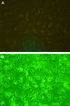
Binding of human class Pi glutathione‐S‐transferase with MDA‐MB‐435S. (A) (100×) MDA‐MB‐435S cells were directly bound with anti‐GST‐fluorescein, washed, and then observed under fluorescence microscope. No fluorescence was emitted from the cells. (B) (100×) When MDA‐MB‐435S cells were incubated with human class Pi glutathione‐S‐transferase followed by anti‐GST‐fluorescein, strong fluorescence could be observed.

References

    1. Mannervik B., Danielson U.H.. Glutathione transferases: structure and catalytic activity. CRC Crit Rev Biochem. 1988; 23: 283–337. - PubMed
    1. Andujar‐Sanchez M., Smith A.W., Clemente‐Jimenez J.M., Rodriguez‐Vico F., Heras‐Vazquez F.J.L., VJara‐Pérez V., et al. Crystallographic and thermodynamic analysis of the binding of S‐octylglutathione to the Tyr 7 to Phe mutant of glutathione S‐transferase from Schistosoma japonicum . Biochemistry. 2005; 44: 1174–1183. - PubMed
    1. Joshua J., Schuster E.F.M., Blanc E., Piper M.D., Thomas J.H., Patel D.S.. Evolutionary conservation of regulated longevity assurance mechanisms. Genome Biol. 2007; 8: 132–137. - PMC - PubMed
    1. Armstrong R.N.. Structure, catalytic mechanism, and evolution of the glutathione transferases. Chem Res Toxicol. 1997; 10: 2–18. - PubMed
    1. Sheehan D., Meade G., Foley V.M., Dowd C.A.. Structure, function, and evolution of glutathione transferases: implications for classification of non‐mammalian members of an ancient enzyme superfamily. Biochem J. 2001; 360: 1–16. - PMC - PubMed

Publication types