. 2011 Nov;27(11):485-93.
doi: 10.1016/j.kjms.2011.06.011.

Dorsal hippocampal N-methyl-D-aspartate glutamatergic and δ-opioidergic systems modulate anxiety behaviors in rats in a noninteractive manner

Affiliations

Dorsal hippocampal N-methyl-D-aspartate glutamatergic and δ-opioidergic systems modulate anxiety behaviors in rats in a noninteractive manner

Jalal Solati. Kaohsiung J Med Sci. 2011 Nov.

Abstract

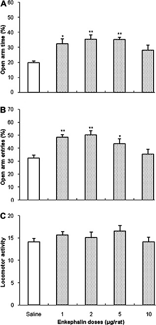
The present study aimed to investigate the effects of N-methyl-d-aspartate (NMDA)-type glutamate receptor agonist, NMDA, on anxiety-like behavior induced by δ-opioid receptor agents in rats, using the elevated plus maze instrument. The dorsal hippocampus (CA1) is known to play an important role in anxiety formation and modulation. Bilateral administration of different doses of δ-opioid receptor agonist, [d-pen2,5] enkephalin acetate hydrate (1 μg/rat, 2 μg/rat, 5 μg/rat, and 10 μg/rat; 1 μL/rat; 0.5 μL/rat in each side), into CA1 area induced an anxiolytic-like effect, demonstrated by substantial increases in the percent of open arm time (OAT%) and percent of open arm entries (OAE%). Intra-CA1 injection of different doses of δ-opioid receptor antagonist, naltrindole hydrochloride (0.25 μg/rat, 0.5 μg/rat, 1 μg/rat, and 2 μg/rat), produced significant anxiogenic-like behavior. Furthermore, intra-CA1 administration of NMDA glutamate receptor agonist, NMDA (0.125 μg/rat, 0.25 μg/rat, 0.5 μg/rat, and 0.75 μg/rat), increased the OAT% and OAE%, indicating anxiolytic-like behavior. However, administration of different doses of NMDA glutamatergic antagonist, MK801 (0.125 μg/rat, 0.25 μg/rat, 0.5 μg/rat, and 1μg/rat), showed no significant effect on the OAT% but decreased the OAE% significantly. The ineffective dose of NMDA (0.125 μg/rat), when coadministered with enkephalin (1 μg/rat, 2 μg/rat, 5 μg/rat, and 10 μg/rat), did not decrease the anxiety behavior significantly. An effective dose of NMDA (0.5 μg/rat), in combination with different doses of naltrindole hydrochloride (0.25 μg/rat, 0.5 μg/rat, 1 μg/rat, and 2 μg/rat), demonstrated no significant interaction with the OAT%, OAE%, and locomotor activity. These results suggest that CA1 δ-opioid and NMDA glutamatergic systems modulate anxiety behaviors in a noninteractive manner.


Figures

Figure 1
Figure 1
A sample of the drug injection site in dorsal hippocampus (CA1). The sections were taken through the brain areas of cannulae placements, and the cannulae placements were verified using the atlas of Paxinos.
Figure 2
Figure 2
Effects of bilateral intra‐CA1 injection of NMDA in the elevated plus maze. Rats were treated with either saline (1 μL/rat) or with NMDA (0.125 μg/rat, 0.25 μg/rat, 0.5 μg/rat, and 0.75 μg/rat). Each bar is mean  ±  standard error of the mean of percent of open arm time (A), percent of open arm entries (B), or locomotor activity (C)—n  =  7. *p  <  0.05, **p  <  0.01, and ***p  <  0.001 when compared with the saline‐treated rats. CA1  =  dorsal hippocampus region; NMDA  =  N‐methyl‐d‐aspartate.
Figure 3
Figure 3
Effects of bilateral intra‐CA1 injection of MK801 in the elevated plus maze. Rats were treated with either saline (1 μL/rat) or with MK801 (0.125 μg/rat, 0.25 μg/rat, 0.5 μg/rat, and 1 μg/rat). Each bar is mean  ±  standard error of the mean of percent of open arm time (A), percent of open arm entries(B), or locomotor activity (C)—n  =  7. *p  <  0.05, when compared with the saline‐treated rats. CA1  =  dorsal hippocampus region.
Figure 4
Figure 4
Effects of bilateral intra‐CA1 injection of enkephalin in the elevated plus maze. Rats were treated with either saline (1 μL/rat) or with enkephalin (1 μg/rat, 2 μg/rat, 5 μg/rat, and 10 μg/rat). Each bar is mean  ±  standard error of the mean of percent of open arm time (A), percent of open arm entries (B), or locomotor activity (C)—n  =  7. *p  <  0.05 and **p  <  0.01, when compared with the saline‐treated rats. CA1  =  dorsal hippocampus region.
Figure 5
Figure 5
Effects of bilateral intra‐CA1 injection of naltrindole in the elevated plus maze. Rats were treated with either saline (1 μL/rat) or with naltrindole (0.25 μg/rat, 0.5 μg/rat, 1 μg/rat, and 2 μg/rat). Each bar is mean  ±  standard error of the mean of percent of open arm time (A), percent of open arm entries (B), or locomotor activity (C)—n  =  7. *p < 0.05 and ** p < 0.01, when compared with the saline‐treated rats. CA1  =  dorsal hippocampus region.
Figure 6
Figure 6
The effect of bilateral injection of enkephalin in the presence or absence of NMDA in the elevated plus maze. Rats were injected saline (1 μL/rat—intra‐CA1) or enkephalin (1 μg/rat, 2 μg/rat, 5 μg/rat, and 10 μg/rat—intra‐CA1) 3 minutes after injection of either saline (1 μL/rat—intra‐CA1) or NMDA (0.125 μg/rat—intra‐CA1). Each bar is mean  ±  standard error of the mean of percent of open arm time (A), percent of open arm entries (B), or locomotor activity (C)—n  =  7. *p  <  0.05, **p  <  0.01, and ***p  <  0.001 when compared with the saline‐treated rats. +p  <  0.05 and ++p  <  0.01 when compared with the saline‐ and NMDA‐treated rats. CA1  =  dorsal hippocampus region; NMDA  =  N‐methyl‐d‐aspartate.
Figure 7
Figure 7
The effect of bilateral injection of naltrindole in the presence or absence of NMDA in the elevated plus maze. Rats were injected saline (1 μL/rat—intra‐CA1) or naltrindole (0.25 μg/rat, 0.5 μg/rat, 1 μg/rat, and 2 μg/rat—intra‐CA1) 3 minutes after injection of either saline (1 μL/rat—intra‐CA1) or NMDA (0.5 μg/rat—intra‐CA1). Each bar is mean  ±  standard error of the mean of percent of open arm time (A), percent of open arm entries (B), or locomotor activity (C)—n  =  7. *p  <  0.05, **p  <  0.01, and ***p  <  0.001 when compared with the saline‐treated rats. +p  <  0.05 when compared with the saline‐ and NMDA‐treated rats. CA1  =  dorsal hippocampus region; NMDA  =  N‐methyl‐d‐aspartate.

Similar articles

Cited by

References

    1. Daumas S., Halley H., Francés B., Lassalle J.. Encoding, consolidation, and retrieval of contextual memory: differential involvement of dorsal CA3 and CA1 hippocampal subregions. Learn Mem. 2005; 12: 375. - PMC - PubMed
    1. File S., Kenny P., Cheeta S.. The role of the dorsal hippocampal serotonergic and cholinergic systems in the modulation of anxiety. Pharmacol Biochem Behav. 2000; 66: 65–72. - PubMed
    1. Ozawa S., Kamiya H., Tsuzuki K.. Glutamate receptors in the mammalian central nervous system. Prog Neurobiol. 1998; 54: 581–618. - PubMed
    1. Guimarães F., Carobrez A., Aguiar J., Graeff F.. Anxiolytic effect in the elevated plus‐maze of the NMDA receptor antagonist AP7 microinjected into the dorsal periaqueductal grey. Psychopharmacology. 1991; 103: 91–94. - PubMed
    1. Zarrindast M., Babapoor‐Farrokhran S., Rezayof A.. Involvement of opioidergic system of the ventral hippocampus, the nucleus accumbens or the central amygdala in anxiety‐related behavior. Life Sci. 2008; 6: 23–24. - PubMed

MeSH terms