. 2011 Dec;219(2):492-8.
doi: 10.1016/j.atherosclerosis.2011.09.038. Epub 2011 Oct 2.

FoxO4 inhibits atherosclerosis through its function in bone marrow derived cells

Affiliations

FoxO4 inhibits atherosclerosis through its function in bone marrow derived cells

Min Zhu et al. Atherosclerosis. 2011 Dec.

Abstract

Objectives: FoxO proteins are transcription factors involved in varieties of cellular processes, including immune cell homeostasis, cytokine production, anti-oxidative stress, and cell proliferation and differentiation. Although these processes are implicated in the development of atherosclerosis, very little is known about the role of FoxO proteins in the context of atherosclerosis. Our objectives were to determine whether and how inactivation of Foxo4, a member of the FoxO family, in vivo promotes atherosclerosis.

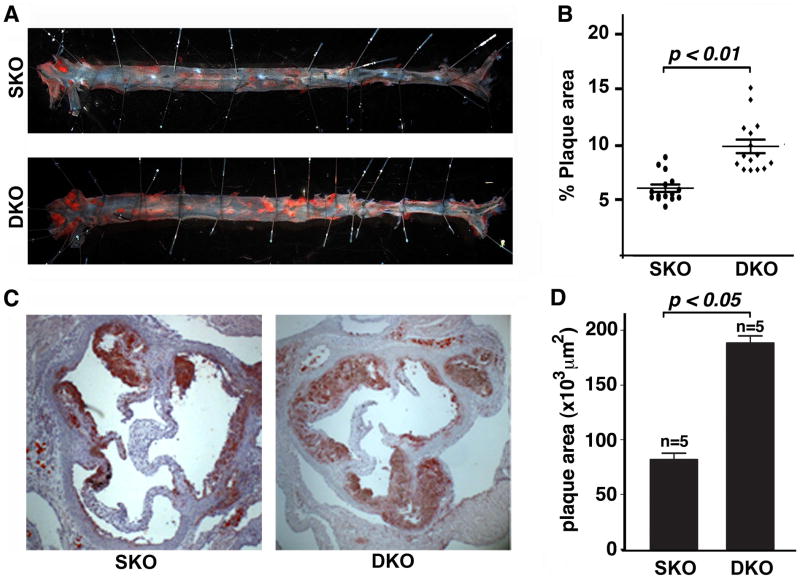
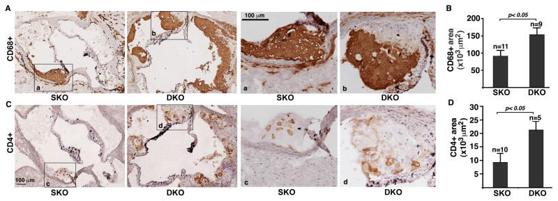
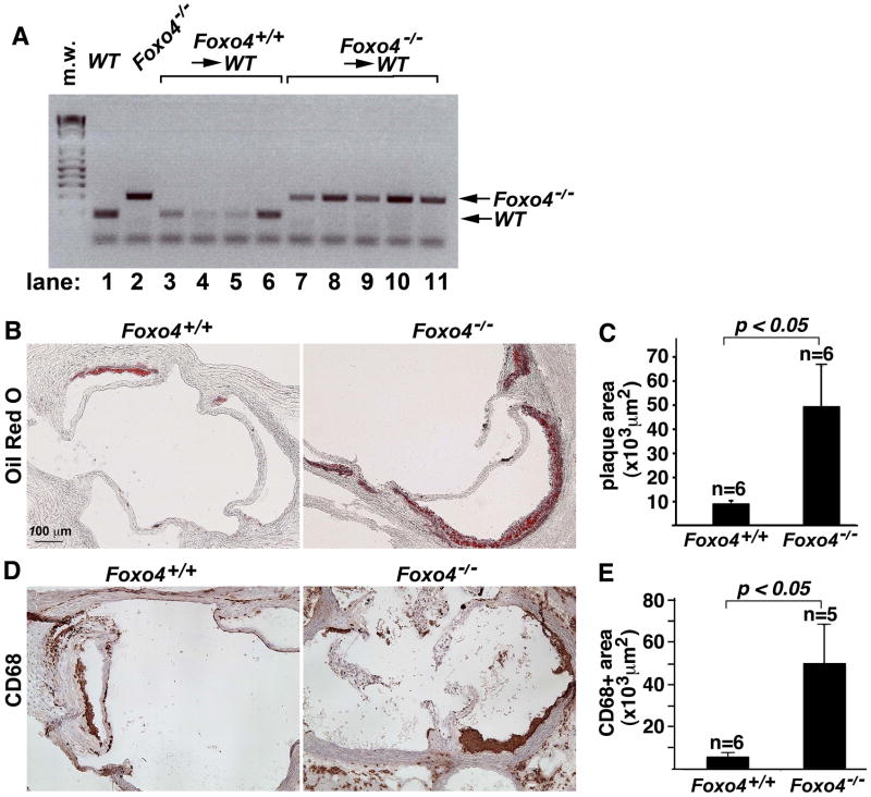
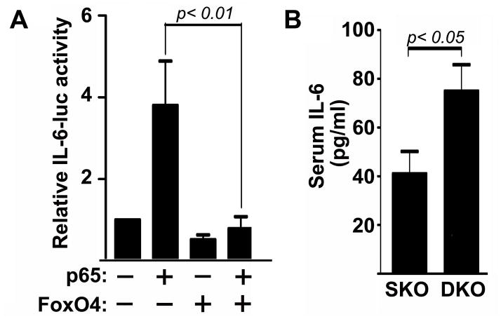
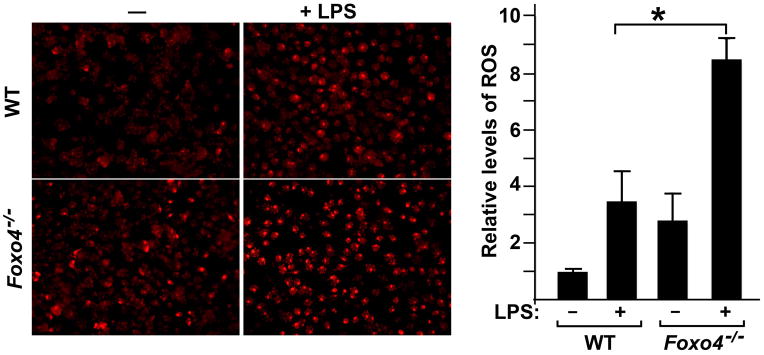
Methods and results: Apolipoprotein E-deficient (apoE(-/-)) mice were crossbred with animals lacking Foxo4 (Foxo4(-/-)). After 10 weeks on a high fat diet (HFD), Foxo4(-/-)apoE(-/-) mice showed elevated atherosclerosis and increased amount of macrophages and T cells in the plaque compared to apoE(-/-) mice. Bone marrow transplantations of chimeric C57B/6 mice reconstituted with either wild-type or Foxo4(-/-) bone marrows indicate that Foxo4-deficiency in bone marrow derived cells sufficiently promoted atherosclerosis. Foxo4-null macrophages produced elevated inflammatory cytokine IL-6 and levels of reactive oxygen species (ROS) in response to lipopolysaccharides in vitro. Serum levels of IL-6 were upregulated in HFD-fed Foxo4(-/-)apoE(-/-) mice compared to those of apoE(-/-) mice.

Conclusions: FoxO4 inhibits atherosclerosis through bone marrow derived cells, possibly by inhibition of ROS and inflammatory cytokines that promote monocyte recruitment and/or retention.


Conflict of interest statement

Conflict of interest: The authors have declared that no conflict of interest exists.

Figures

Figure 1
Figure 1. Inactivation of Foxo4 promotes atherosclerosis formation
(A)en face analysis of atherosclerotic lesions of apoE SKO and Foxo4/apoE DKO mice after 10 weeks of HFD. The aorta was removed in its entirety and opened longitudinally. The artery was pinned on a surface and stained with oil red O. (B) The area of intima that is covered by lesions was outlined and determined as a percentage of total intimal surface area. Foxo4/apoE DKO mice have significantly higher amounts of atherosclerotic lesions than apoE SKO mice (n=15, mean ± SEM, p < 0.01). (C) Representative photomicrographs of atherosclerotic plaques at the aortic root of a Foxo4/apoE DKO and apoE SKO mouse. Frozen cross-sections of the aortic root were obtained and stained with oil red O. (D) Cross-sectional plaque areas were traced manually and measured morphometrically. Foxo4/apoE DKO mice have significantly higher amounts of atherosclerotic lesions than apoE SKO (n=5, mean ± SEM, p < 0.05).
Figure 2
Figure 2. Macrophages and T cells are upregulated in the atherosclerotic plaques of Foxo4/apoE DKO mice
Immunohistochemistry was performed on sections of the aortic root of HFD-fed Foxo4/apoE DKO and apoE SKO mice using antibodies against CD68 (A and B) ad CD4 (C and D). (A) Representative of photomicrograph of sections stained with anti-CD68 antibody. High-magnification images of the insets (a and b) on the left panels are shown on the right panels. (B) CD68+ stains in DKO mice were upregulated compared to that of control SKO mice. Values are mean ± SEM, p < 0.05. High-magnification images of the insets (c and d) on the left panels are shown in the right panels. (C) Representative of photomicrograph of sections stained with anti-CD4 antibody. (D) CD4+ T cells in the lesions of DKO were significantly upregulated compared to control SKO mice. Values are mean ± SEM, p < 0.05.
Fig. 3
Fig. 3. Foxo4-deficient BMs are sufficient to promote enhanced atherosclerosis
Female WT C57B/6 mice were irradiated one day before the BMT. The BM suspension (2×10⁶) from either control WT or Foxo4−/− mice was injected intravenously via the tail vein. After 4 weeks recovery, the chimeric mice were fed HFD for four months and sacrificed. (A) PCR genotyping of bone marrows of wild type (lane 1), Foxo4-null (lane 2), and chimeric mice received with Foxo4+/+ bone marrows (lanes, 3–6), and Foxo4−/− bone marrows (lanes, 7–11). The peripheral blood DNA from Foxo4−/−→WT mice has the presence of donor Foxo4−/− genotype with negligible host DNA remaining. (B) Representative photographs of plaques in the aortic root of chimeric mice received with Foxo4-null (left) and wild type bone marrows. (C) The atherosclerotic lesion in chimeric mice reconstituted with Foxo4−/− BMs is significantly larger than that of mice received with wild type BMs. Values are mean ± SEM, p <0.05. (D) Macrophage contents in the lesions of chimeric mice were quantified using anti-CD68 antibody. (E) Chimeric mice transplanted with Foxo4-null bone marrows have higher number of macrophages than those received with wild type bone marrows after fed with HFD for 4 months. Values are mean ± SEM, p < 0.05.
Figure 4
Figure 4. FoxO4 inhibits IL-6 expression
(A) The IL-6-luciferase reporter was transfected into macrophages in the presence of the plasmids indicated. Luciferase activities were measured 24 hrs after transfection and normalized against co-transfected β-galactosidase. FoxO4 inhibited NF-κB activated IL-6-luc activity (n=4, mean ± SEM). (B) The serum levels of IL-6 in HFD-fed Foxo4/apoE DKO and apoE SKO mice were quantified with a standard IL-6 ELISA assay (n=15, mean ± SEM).
Figure 5
Figure 5. Inactivation of Foxo4 in macrophages leads to enhanced production of ROS in response to LPS
Equal numbers of peritoneal macrophages from wild type and Foxo4-null mice were plated onto 6-well plates in triplicates, stimulated with or without LPS. Cells were stained with DHE. Levels of ROS were measured as the product of the intensity and area of DHE-positive cells and expressed relative to the value of WT cells without LPS stimulation. n=4±SEM, *, p < 0.05.

Similar articles

Cited by

References

    1. Andersson J, Libby P, Hansson GK. Adaptive immunity and atherosclerosis. Clin Immunol. 2010;134:33–46. - PubMed
    1. Ando H, Tsuruoka S, Yamamoto H, Takamura T, Kaneko S, Fujimura A. Regulation of cholesterol 7alpha-hydroxylase mRNA expression in C57BL/6 mice fed an atherogenic diet. Atherosclerosis. 2005;178:265–269. - PubMed
    1. Aparicio-Vergara M, Shiri-Sverdlov R, de Haan G, Hofker MH. Bone marrow transplantation in mice as a tool for studying the role of hematopoietic cells in metabolic and cardiovascular diseases. Atherosclerosis. 2010 [Epub ahead of print] - PubMed
    1. Baglione J, Smith JD. Quantitative assay for mouse atherosclerosis in the aortic root. Methods Mol Med. 2006;129:83–95. - PubMed
    1. Brasier AR. The nuclear factor-kappaB-interleukin-6 signalling pathway mediating vascular inflammation. Cardiovasc Res. 2010;86:211–8. - PMC - PubMed

Publication types

MeSH terms