Review
. 2011 Oct 4:13:e31.
doi: 10.1017/S1462399411002018.

Interferon-gamma- and perforin-mediated immune responses for resistance against Toxoplasma gondii in the brain

Affiliations
Review

Interferon-gamma- and perforin-mediated immune responses for resistance against Toxoplasma gondii in the brain

Yasuhiro Suzuki et al. Expert Rev Mol Med. .

Abstract

Toxoplasma gondii is an obligate intracellular protozoan parasite that causes various diseases, including lymphadenitis, congenital infection of fetuses and life-threatening toxoplasmic encephalitis in immunocompromised individuals. Interferon-gamma (IFN-γ)-mediated immune responses are essential for controlling tachyzoite proliferation during both acute acquired infection and reactivation of infection in the brain. Both CD4+ and CD8+ T cells produce this cytokine in response to infection, although the latter has more potent protective activity. IFN-γ can activate microglia, astrocytes and macrophages, and these activated cells control the proliferation of tachyzoites using different molecules, depending on cell type and host species. IFN-γ also has a crucial role in the recruitment of T cells into the brain after infection by inducing expression of the adhesion molecule VCAM-1 on cerebrovascular endothelial cells, and chemokines such as CXCL9, CXCL10 and CCL5. A recent study showed that CD8+ T cells are able to remove T. gondii cysts, which represent the stage of the parasite in chronic infection, from the brain through their perforin-mediated activity. Thus, the resistance to cerebral infection with T. gondii requires a coordinated network using both IFN-γ- and perforin-mediated immune responses. Elucidating how these two protective mechanisms function and collaborate in the brain against T. gondii will be crucial in developing a new method to prevent and eradicate this parasitic infection.


Figures
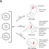
Fig. 1
Fig. 1
Schematic figures of effector mechanisms mediated by IFN-γ and perforin in resistance against T. gondii in the brain. Panel A. IFN-γ-mediated effector mechanisms against tachyzoites in the brain Microglia: Microglia activated by IFN-γ plus lipopolysaccharide inhibit proliferation of tachyzoites through nitric oxide (NO) production by inducible nitric oxide synthase (NOS2). An involvement of both NO-dependent and –independent mechanisms is suggested in the resistance of murine microglia activated by a combination of IFN-γ and TNF-α. The mechanisms of the NO-independent inhibitory effect is unknown. Astrocytes: Murine astrocytes activated by IFN-γ inhibit proliferation of tachyzoites by Irgm3 (IGTP), one of p47 GTPases. TNF-α and IFN-γ synergistically activate indoleamine 2,3-dioxygenase in human glioblastoma cell lines and native astrocytes, and this IDO activity results in a strong toxoplasmostatic effect in these activated cells. Macrophages: NO production by NOS2 was first shown to be a mechanism of macrophages activated by IFN-γ to restrict tachyzoite growth in vitro. However, recent studies indicated that p47 GTPases such as IGTP and IIGP1 are critical for the activity of activated macrophages to restrict tachyzoite growth. Studies using peritoneal macrophages from mice primed with live attenuated tachyzoites showed that the IFN-γ-induced, IGTP-mediated killing was preceded by parasitophorous vacuole (the vacuole containing tachyzoites) indentation, vesiculation, disruption, and stripping of the parasite plasma membrane. Denuded parasites are then enveloped in autophagosome-like vacuoles, which ultimately fuse with lysosomes. In peritoneal macrophages activated in vitro by IFN-γ plus LPS, autophagy protein Atg5 is required for recruitment of IIGP1 to the parasitophorous vacuole membrane and parasite clearance. Panel B. IFN-γ-independent effector mechanisms against tachyzoites in the brain Microglia: CD40-CD40L interaction induces autophagy-mediated control of tachyzoites within microglia. Microglia from mice lacking the autophagy molecules, Beclin 1 or Atg7, failed to kill intracellular tachyzoites in vitro after stimulation through CD40. Macrophages: P2X7 receptor activation by extracellular ATP kills tachyzoites in infected macrophages, and this killing occurs in parallel with host cell apoptosis. Although this killing mechanism itself does not require IFN-γ, P2X7 receptor expression can be upregulated by this cytokine. Panel C. Perforin-mediated effector mechanisms against tachyzoites in the brain Tissue cysts of T. gondii are formed mostly within neurons and astrocytes in the brain. Immune T cells can eliminate the cysts from the brain when the T cells are transferred into infected immunodeficient animals that have already developed large numbers of the cysts. The T cell-mediated immune process was associated with accumulation of microglia and macrophages around tissue cysts. Since the accumulated phagocytes penetrate within the cyst, these cells appear be the main effector cells that destroy the cysts and eliminate them from the brain after initiation of this process by immune T cells. CD8+ immune T cells have a potent activity to remove the cysts, and their anti-cyst activity requries perforin. Perforin is the major molecule that mediates cytolysis of target cells by CD8+ cytotoxic T cells. Therefore, CD8+ T cells appear to induce elimination of T. gondii cysts through their perforin-mediated cytotoxic activity.
Fig. 1
Fig. 1
Schematic figures of effector mechanisms mediated by IFN-γ and perforin in resistance against T. gondii in the brain. Panel A. IFN-γ-mediated effector mechanisms against tachyzoites in the brain Microglia: Microglia activated by IFN-γ plus lipopolysaccharide inhibit proliferation of tachyzoites through nitric oxide (NO) production by inducible nitric oxide synthase (NOS2). An involvement of both NO-dependent and –independent mechanisms is suggested in the resistance of murine microglia activated by a combination of IFN-γ and TNF-α. The mechanisms of the NO-independent inhibitory effect is unknown. Astrocytes: Murine astrocytes activated by IFN-γ inhibit proliferation of tachyzoites by Irgm3 (IGTP), one of p47 GTPases. TNF-α and IFN-γ synergistically activate indoleamine 2,3-dioxygenase in human glioblastoma cell lines and native astrocytes, and this IDO activity results in a strong toxoplasmostatic effect in these activated cells. Macrophages: NO production by NOS2 was first shown to be a mechanism of macrophages activated by IFN-γ to restrict tachyzoite growth in vitro. However, recent studies indicated that p47 GTPases such as IGTP and IIGP1 are critical for the activity of activated macrophages to restrict tachyzoite growth. Studies using peritoneal macrophages from mice primed with live attenuated tachyzoites showed that the IFN-γ-induced, IGTP-mediated killing was preceded by parasitophorous vacuole (the vacuole containing tachyzoites) indentation, vesiculation, disruption, and stripping of the parasite plasma membrane. Denuded parasites are then enveloped in autophagosome-like vacuoles, which ultimately fuse with lysosomes. In peritoneal macrophages activated in vitro by IFN-γ plus LPS, autophagy protein Atg5 is required for recruitment of IIGP1 to the parasitophorous vacuole membrane and parasite clearance. Panel B. IFN-γ-independent effector mechanisms against tachyzoites in the brain Microglia: CD40-CD40L interaction induces autophagy-mediated control of tachyzoites within microglia. Microglia from mice lacking the autophagy molecules, Beclin 1 or Atg7, failed to kill intracellular tachyzoites in vitro after stimulation through CD40. Macrophages: P2X7 receptor activation by extracellular ATP kills tachyzoites in infected macrophages, and this killing occurs in parallel with host cell apoptosis. Although this killing mechanism itself does not require IFN-γ, P2X7 receptor expression can be upregulated by this cytokine. Panel C. Perforin-mediated effector mechanisms against tachyzoites in the brain Tissue cysts of T. gondii are formed mostly within neurons and astrocytes in the brain. Immune T cells can eliminate the cysts from the brain when the T cells are transferred into infected immunodeficient animals that have already developed large numbers of the cysts. The T cell-mediated immune process was associated with accumulation of microglia and macrophages around tissue cysts. Since the accumulated phagocytes penetrate within the cyst, these cells appear be the main effector cells that destroy the cysts and eliminate them from the brain after initiation of this process by immune T cells. CD8+ immune T cells have a potent activity to remove the cysts, and their anti-cyst activity requries perforin. Perforin is the major molecule that mediates cytolysis of target cells by CD8+ cytotoxic T cells. Therefore, CD8+ T cells appear to induce elimination of T. gondii cysts through their perforin-mediated cytotoxic activity.

Similar articles

Cited by

References

    1. McCabe RE, Remington JS. Toxoplasma gondii. In: Mandell GL, Douglas RG, Bennett JE, editors. Principles and Practice of Infectious Diseases. Churchill Livingstone Inc.; New York: 1990. 2090.
    1. Suzuki Y, Orellana MA, Schreiber RD, Remington JS. Interferon gamma: the major mediator of resistance against Toxoplasma gondii. Science. 1988;240:516–518. - PubMed
    1. Denkers EY, Gazzinelli RT. Regulation and function of Tcellmediated immunity during Toxoplasma gondii infection. Clin.Microbiol.Rev. 1998;11:569–588. - PMC - PubMed
    1. Boyer K, Marcinak J, McLeod R. Toxoplasma gondii (Toxoplasmosis) In: Long S, Pickering LK, Prober CG, editors. In Principles and Practice of Pediatric Infectious Diseases. 3rd edition Churchill Livingstone; New York: 2007.
    1. Israelski DM, Remington JS. Toxoplasmosis in the non-AIDS immunocompromised host. In: Remington JS, Swrltz M, editors. Curr.Clin.Top.Infect.Dis. Blackwell Scientific Publications; London: 1993. pp. 322–356. - PubMed

Publication types

MeSH terms