

Review
doi: 10.1016/j.abb.2011.09.012.
Epub 2011 Oct 7.
Allosteric regulation of phenylalanine hydroxylase
Affiliations
- PMID: 22005392
- PMCID: PMC3271142
- DOI: 10.1016/j.abb.2011.09.012
Item in Clipboard
Review
Allosteric regulation of phenylalanine hydroxylase
Arch Biochem Biophys.
.
Abstract
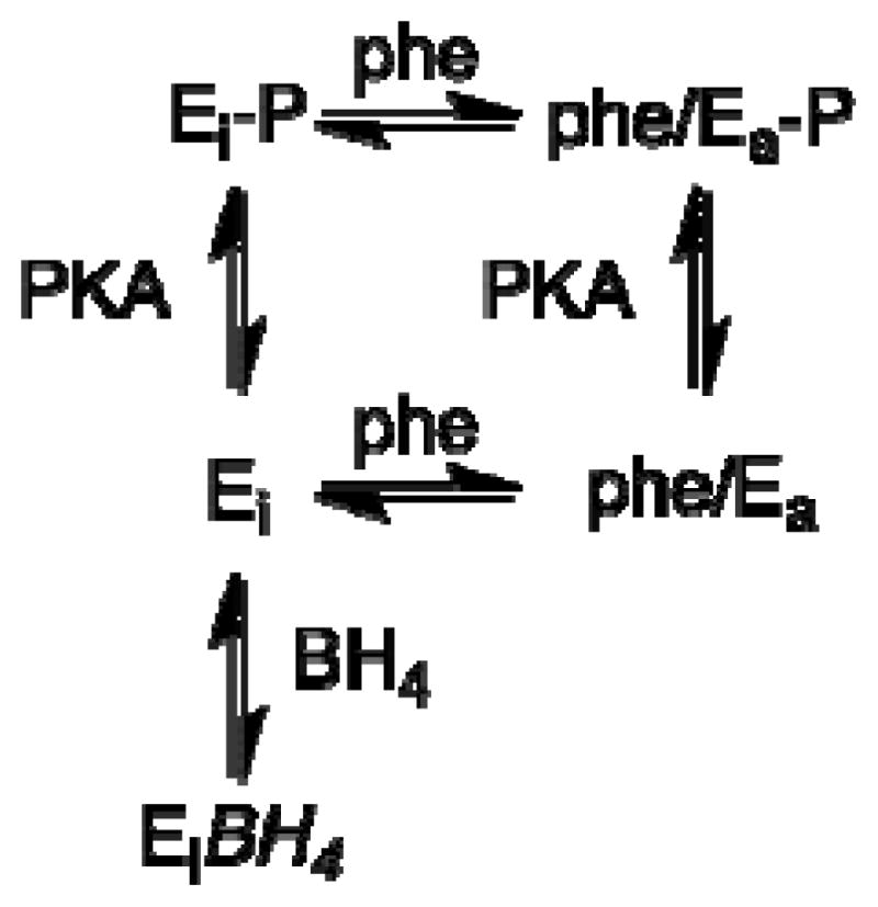
The liver enzyme phenylalanine hydroxylase is responsible for conversion of excess phenylalanine in the diet to tyrosine. Phenylalanine hydroxylase is activated by phenylalanine; this activation is inhibited by the physiological reducing substrate tetrahydrobiopterin. Phosphorylation of Ser16 lowers the concentration of phenylalanine for activation. This review discusses the present understanding of the molecular details of the allosteric regulation of the enzyme.
Copyright © 2011 Elsevier Inc. All rights reserved.
Figures

Structure of PheH. A, Structure of the intact tetramer; B, structure of a dimer with the regulatory and catalytic domains on the same subunit colored different shades of red or blue; C, the structure of an individual monomer with the regulatory domain (blue) in cartoon mode and the active site iron shown as an orange sphere. Figures 1A and 1B were constructed by overlaying the dimeric structure of rat PheH lacking the C-terminal helix (pdb file 2PHM) and the tetrameric structure of human PheH lacking the regulatory domain (pdb file 2PAH). Figure C was constructed using pdb file 2PHM.

Potential allosteric sites in the regulatory domain of PheH. Orange: binding site for BH4 based on structure of PCD/DCoH. Magenta: residues 42–48 and 63–69, potential binding sites for phenylalanine. Green: residues 19–31. Blue: residues 32–116. Red: catalytic domain, residues 117–426. The figure is based on pdb file 2PHM.

Proposed interactions [96] between Ser23 in the regulatory domain and BH4 in the active site of the binary BH4 complex (magenta carbons and blue H-bonds) and the ternary BH4-amino acid complex (green carbons and H-bonds).


References
-
- Fitzpatrick PF. Ann Rev Biochem. 1999;68:355–381. - PubMed
-
- Almas B, Le Bourdelles B, Flatmark T, Mallet J, Haavik J. Eur J Biochem. 1992;209:249–255. - PubMed
-
- Ramsey AJ, Fitzpatrick PF. Biochemistry. 1998;37:8980–8986. - PubMed
-
- McKinney J, Knappskog PM, Haavik J. J Neurochem. 2005;92:311–320. - PubMed
Publication types
MeSH terms
Substances
Grants and funding
LinkOut - more resources
Full Text Sources
Molecular Biology Databases
