

Derivation of smooth muscle cells with neural crest origin from human induced pluripotent stem cells
- PMID: 22005509
- PMCID: PMC3257812
- DOI: 10.1159/000331412
Derivation of smooth muscle cells with neural crest origin from human induced pluripotent stem cells
Abstract
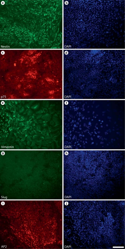
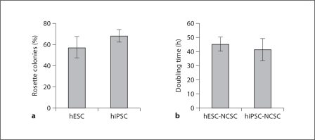
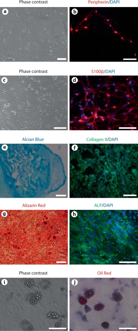
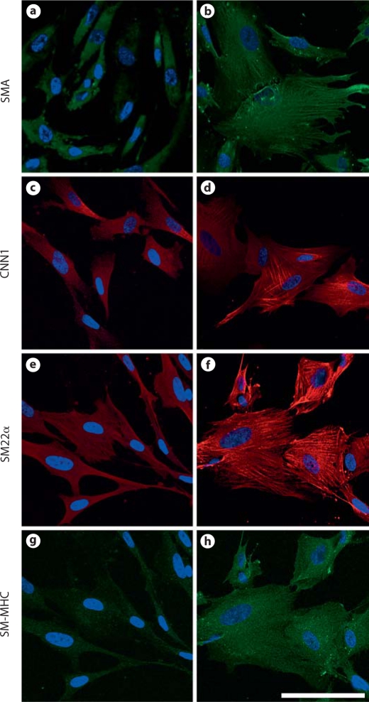
The heterogeneity of vascular smooth muscle cells (SMCs) is related to their different developmental origins such as the neural crest and mesoderm. Derivation of SMCs from different origins will provide valuable in vitro models for the investigation of vascular development and diseases. From the perspective of regenerative medicine and tissue engineering, an expandable cell source of SMCs is required for the construction of tissue-engineered blood vessels. In this study, we developed a robust protocol to derive neural crest stem cells (NCSCs) from human embryonic stem cells (ESCs) and induced pluripotent stem cells (iPSCs). NCSCs derived from ESCs and iPSCs were expandable with similar cell doubling times. NCSCs were capable of differentiating into neural and mesenchymal lineages. TGF-β1 induced the expression of SMC markers calponin-1, SM22α, and smooth muscle myosin heavy chain and resulted in the assembly of smooth muscle α-actin, calponin-1, and SM22α into stress fibers. This work provides a basis for using iPSCs to study SMC biology and deriving vascular cells for tissue engineering.
Copyright © 2011 S. Karger AG, Basel.
Figures


Similar articles
-
Uniaxial mechanical strain modulates the differentiation of neural crest stem cells into smooth muscle lineage on micropatterned surfaces.PLoS One. 2011;6(10):e26029. doi: 10.1371/journal.pone.0026029. Epub 2011 Oct 7. PLoS One. 2011. PMID: 22016804 Free PMC article.
-
Differentiation of Neural Crest Stem Cells in Response to Matrix Stiffness and TGF-β1 in Vascular Regeneration.Stem Cells Dev. 2020 Feb 15;29(4):249-256. doi: 10.1089/scd.2019.0161. Epub 2019 Dec 17. Stem Cells Dev. 2020. PMID: 31701817
-
Differentiation defect in neural crest-derived smooth muscle cells in patients with aortopathy associated with bicuspid aortic valves.EBioMedicine. 2016 Aug;10:282-90. doi: 10.1016/j.ebiom.2016.06.045. Epub 2016 Jul 1. EBioMedicine. 2016. PMID: 27394642 Free PMC article.
-
Embryonic origins of human vascular smooth muscle cells: implications for in vitro modeling and clinical application.Cell Mol Life Sci. 2014 Jun;71(12):2271-88. doi: 10.1007/s00018-013-1554-3. Epub 2014 Jan 18. Cell Mol Life Sci. 2014. PMID: 24442477 Free PMC article. Review.
-
Pluripotent stem cell differentiation into vascular cells: a novel technology with promises for vascular re(generation).Pharmacol Ther. 2011 Jan;129(1):29-49. doi: 10.1016/j.pharmthera.2010.10.004. Epub 2010 Oct 20. Pharmacol Ther. 2011. PMID: 20965210 Review.
Cited by
-
Activation of Hes1 and Msx1 in Transgenic Mouse Embryonic Stem Cells Increases Differentiation into Neural Crest Derivatives.Int J Mol Sci. 2018 Dec 13;19(12):4025. doi: 10.3390/ijms19124025. Int J Mol Sci. 2018. PMID: 30551562 Free PMC article.
-
Small-diameter vascular graft engineered using human embryonic stem cell-derived mesenchymal cells.Tissue Eng Part A. 2014 Feb;20(3-4):740-50. doi: 10.1089/ten.TEA.2012.0738. Tissue Eng Part A. 2014. PMID: 24125588 Free PMC article.
-
Human ectodermal organoids reveal the cellular origin of DiGeorge Syndrome.bioRxiv [Preprint]. 2025 Aug 8:2025.08.08.669417. doi: 10.1101/2025.08.08.669417. bioRxiv. 2025. PMID: 40855884 Free PMC article. Preprint.
-
Differentiation of Brain Pericyte-Like Cells from Human Pluripotent Stem Cell-Derived Neural Crest.Curr Protoc. 2021 Jan;1(1):e21. doi: 10.1002/cpz1.21. Curr Protoc. 2021. PMID: 33484491 Free PMC article.
-
Induced pluripotent stem cell-derived vascular smooth muscle cells.Vasc Biol. 2019 Dec 12;2(1):R1-R15. doi: 10.1530/VB-19-0028. eCollection 2020. Vasc Biol. 2019. PMID: 32923972 Free PMC article. Review.
References
-
- Adam P.J., Regan C.P., Hautmann M.B., Owens G.K. Positive- and negative-acting Kruppel-like transcription factors bind a transforming growth factor β control element required for expression of the smooth muscle cell differentiation marker SM22α in vivo. J Biol Chem. 2000;275:37798–37806. - PubMed
-
- Bergwerff M., Verberne M.E., DeRuiter M.C., Poelmann R.E., Gittenberger-de Groot A.C. Neural crest cell contribution to the developing circulatory system: implications for vascular morphology? Circ Res. 1998;82:221–231. - PubMed
-
- Chen S., Lechleider R.J. Transforming growth factor-β-induced differentiation of smooth muscle from a neural crest stem cell line. Circ Res. 2004;94:1195–1202. - PubMed
-
- Crane J.F., Trainor P.A. Neural crest stem and progenitor cells. Annu Rev Cell Dev Biol. 2006;22:267–286. - PubMed
-
- DeBakey M.E., Glaeser D.H. Patterns of atherosclerosis: effect of risk factors on recurrence and survival-analysis of 11,890 cases with more than 25-year follow-up. Am J Cardiol. 2000;85:1045–1053. - PubMed
Publication types
MeSH terms
Grants and funding
LinkOut - more resources
Full Text Sources
Other Literature Sources
