. 2012;195(1-2):108-21.
doi: 10.1159/000331415. Epub 2011 Oct 14.

A novel ovine ex vivo arteriovenous shunt model to test vascular implantability

Affiliations

A novel ovine ex vivo arteriovenous shunt model to test vascular implantability

Haofan Peng et al. Cells Tissues Organs. 2012.

Abstract

The major objective of successful development of tissue-engineered vascular grafts is long-term in vivo patency. Optimization of matrix, cell source, surface modifications, and physical preconditioning are all elements of attaining a compatible, durable, and functional vascular construct. In vitro model systems are inadequate to test elements of thrombogenicity and vascular dynamic functional properties while in vivo implantation is complicated, labor-intensive, and cost-ineffective. We proposed an ex vivo ovine arteriovenous shunt model in which we can test the patency and physical properties of vascular grafts under physiologic conditions. The pressure, flow rate, and vascular diameter were monitored in real-time in order to evaluate the pulse wave velocity, augmentation index, and dynamic elastic modulus, all indicators of graft stiffness. Carotid arteries, jugular veins, and small intestinal submucosa-based grafts were tested. SIS grafts demonstrated physical properties between those of carotid arteries and jugular veins. Anticoagulation properties of grafts were assessed via scanning electron microscopy imaging, en face immunostaining, and histology. Luminal seeding with endothelial cells greatly decreased the attachment of thrombotic components. This model is also suture free, allowing for multiple samples to be stably processed within one animal. This tunable (pressure, flow, shear) ex vivo shunt model can be used to optimize the implantability and long-term patency of tissue-engineered vascular constructs.


Figures

Fig. 1
Fig. 1
Fabrication of the fibrin-glued SIS vascular graft. Fibrin-glued SIS vascular graft end view (a) and longitudinal view (b). Native ovine carotid artery end view (c) and longitudinal view (d). e H&E-stained cross section of fibrin-glued SIS depicts four layers of SIS adhered with fibrin glue (22.0 mg/ml). Arrows indicate the interfaces between SIS layers. f Illustration of fibrin glue application on SIS. Fibrinogen was homogeneously spread on SIS and thrombin was added with a syringe as the layers were wrapped around the mandrel.
Fig. 2
Fig. 2
The fibrin-glued SIS vascular graft showed increased burst pressure at higher fibrin glue concentrations. Fibrin glue was used at 5.5 mg/ml (n = 4), 11 mg/ml (n = 5), or 22 mg/ml (n = 7). The asterisk denotes statistical significance between the indicated samples and those at the highest fibrin concentration of 22 mg/ml (p < 0.05).
Fig. 3
Fig. 3
Confluence and alignment of endothelial cells on the luminal surface of SIS. BAECs were seeded in the SIS lumen and cultured statically (a, b) or under shear (c, d) for 10 days. e Illustration of the shear ramping protocol: 8 h of seeding under rotation was followed by 7 days of static culture, ramping from 0 to 6 dyne/cm2 within 24 h and keeping the grafts at that shear level for 2 additional days. Scale bars = 50 μm (a, c) and 20 μm (b, d).
Fig. 4
Fig. 4
Ovine arteriovenous shunt model. a Image of the arteriovenous shunt model. b Schematic of the arteriovenous shunt model. The endothelial cell-seeded, fibrin-glued SIS graft was mounted in a bioreactor chamber and connected in series with a pressure transducer, flow transducer, and laser micrometer. A = Carotid artery; PT = pressure transducer; FT = flow rate transducer; C = chamber (grafts); R = restrictor; COM = computer and digital reader; LED = LED micrometer; V = jugular vein.
Fig. 5
Fig. 5
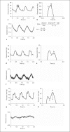
Vascular stiffness measurement using PWV, AIx, and Em. a–c Jugular veins. d–f SIS grafts. g–i Carotid arteries. a, d, g Pressure wave form. b, e, h Magnification of the pressure wave peak is used to calculate the AIx. c, f, i Dynamic diameter change. Ps = Systolic pressure; Pd = diastolic pressure; Pi = inflection point that indicates the beginning upstroke of the reflected pressure wave; TA = time corresponding to the pressure foot (point A); TA′ = time at point A′. PT = Pressure transducer, LED = LED micrometer. For each graft PWV represents the average of 2–3 waveforms. j Quantification of PWV. k Quantification of AIx. l Quantification of Em. The asterisk denotes a significant difference between the indicated sample and SIS (n = 3 grafts). For each graft PWV represents the average of 2–3 waveforms.
Fig. 5
Fig. 5
Vascular stiffness measurement using PWV, AIx, and Em. a–c Jugular veins. d–f SIS grafts. g–i Carotid arteries. a, d, g Pressure wave form. b, e, h Magnification of the pressure wave peak is used to calculate the AIx. c, f, i Dynamic diameter change. Ps = Systolic pressure; Pd = diastolic pressure; Pi = inflection point that indicates the beginning upstroke of the reflected pressure wave; TA = time corresponding to the pressure foot (point A); TA′ = time at point A′. PT = Pressure transducer, LED = LED micrometer. For each graft PWV represents the average of 2–3 waveforms. j Quantification of PWV. k Quantification of AIx. l Quantification of Em. The asterisk denotes a significant difference between the indicated sample and SIS (n = 3 grafts). For each graft PWV represents the average of 2–3 waveforms.
Fig. 6
Fig. 6
SIS grafts seeded with BAECs in static or sheared conditions demonstrated coagulation deposits in gross images and histology. a, f Static BAEC-seeded grafts. b, g Sheared BAEC-seeded grafts. c, h SIS only. d, i Jugular vein. e, j Carotid artery. Scale bars = 1 cm (a–e) and 50 μm (f–h). The arrows point to platelets or red blood cells.
Fig. 7
Fig. 7
The endothelialized surface of grafts exhibited better antithrombogenic properties. Platelets on the surface of grafts were stained with anti-CD61 (green) to identify platelets. Hoechst (blue) and phalloidin Alexa 546 (red). a, b Static culture: some individual platelets. c, d Shear condition: platelets were seen at the intercellular area; e, f SIS only: without endothelial cells large platelet aggregates were observed throughout the lumen of the grafts. The platelet aggregate is circled. g, h Jugular vein: minimum platelet adhesion. i, j Carotid artery: minimum platelet adhesion. Arrows indicate platelets. Scale bars = 50 μm (a, c, e, g, i) and 20 μm (b, d, f, h, j).
Fig. 8
Fig. 8
Endothelialization of the grafts reduced luminal coagulation. SEM images of the graft lumen. Platelets, red blood cells, white blood cells, and fibrin mesh can be clearly visualized and distinguished. Severe coagulation was observed throughout the cell-free surface of SIS. a, b Static. c, d Sheared. e, f SIS only. g, h Jugular vein. i, j Carotid artery. k Quantification of the luminal area that was covered by platelets at the indicated conditions. P = Platelets; W = white blood cells; R = red blood cells; F = fibrin mesh. The asterisk denotes a significant difference as compared to the SIS-only surface. # denotes a significant difference between the indicated grafts (n = 3, p < 0.05).

Similar articles

Cited by

References

    1. Abbott W.M., Megerman J., Hasson J.E., L'Italien G., Warnock D.F. Effect of compliance mismatch on vascular graft patency. J Vasc Surg. 1987;5:376–382. - PubMed
    1. Aoki J., Serruys P.W., van Beusekom H., Ong A.T., McFadden E.P., Sianos G., van der Giessen W.J., Regar E., de Feyter P.J., Davis H.R., Rowland S., Kutryk M.J. Endothelial progenitor cell capture by stents coated with antibody against CD34: the HEALING-FIM (Healthy Endothelial Accelerated Lining Inhibits Neointimal Growth-First In Man) Registry. J Am Coll Cardiol. 2005;45:1574–1579. - PubMed
    1. Baguet J.P., Kingwell B.A., Dart A.L., Shaw J., Ferrier K.E., Jennings G.L. Analysis of the regional pulse wave velocity by Doppler: methodology and reproducibility. J Hum Hypertens. 2003;17:407–412. - PubMed
    1. Brass L.F., Zhu L., Stalker T.J. Novel therapeutic targets at the platelet vascular interface. Arterioscler Thromb Vasc Biol. 2008;28:s43–50. - PubMed
    1. Chin-Quee S.L., Hsu S.H., Nguyen-Ehrenreich K.L., Tai J.T., Abraham G.M., Pacetti S.D., Chan Y.F., Nakazawa G., Kolodgie F.D., Virmani R., Ding N.N., Coleman L.A. Endothelial cell recovery, acute thrombogenicity, and monocyte adhesion and activation on fluorinated copolymer and phosphorylcholine polymer stent coatings. Biomaterials. 2009;31:648–657. - PubMed

Publication types