. 2011 Dec 1;589(Pt 23):5663-76.
doi: 10.1113/jphysiol.2011.215384. Epub 2011 Oct 17.

Selective silencing of Na(V)1.7 decreases excitability and conduction in vagal sensory neurons

Affiliations

Selective silencing of Na(V)1.7 decreases excitability and conduction in vagal sensory neurons

Yukiko Muroi et al. J Physiol. .

Abstract

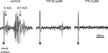
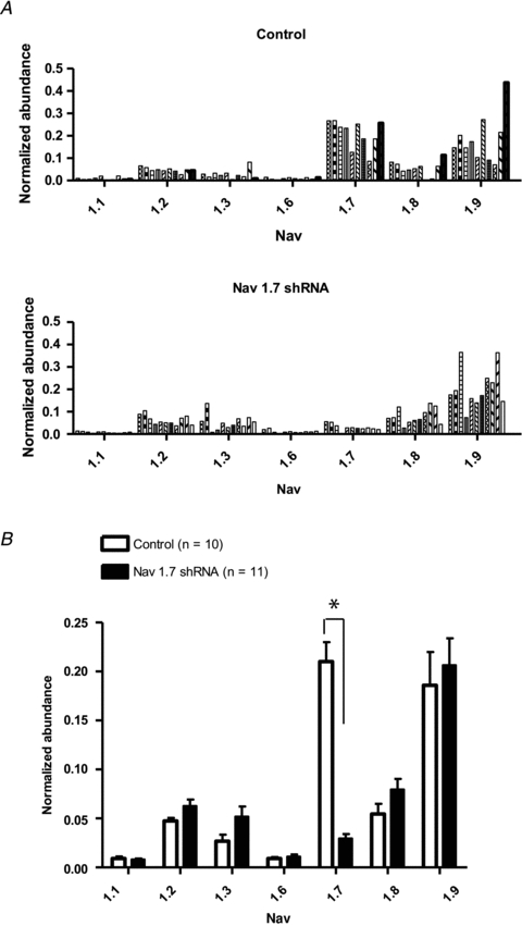
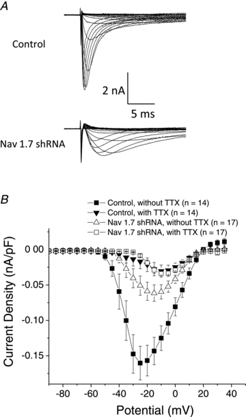
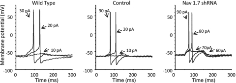
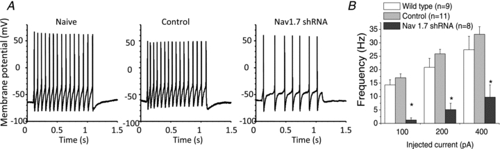
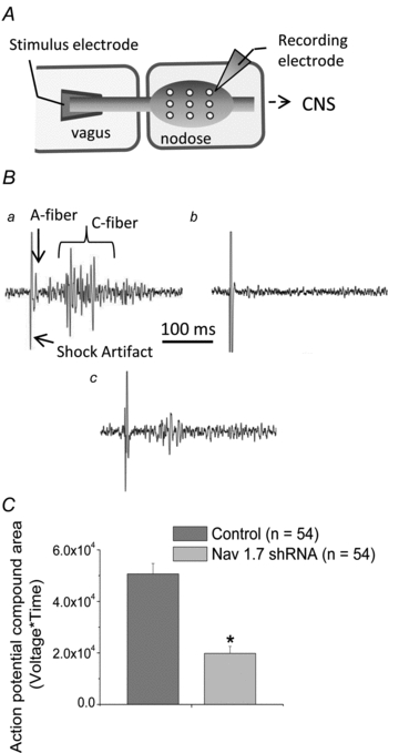
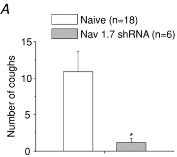
There has been much information learned in recent years about voltage gated sodium channel (Na(V)) subtypes in somatosensory pain signalling, but much less is known about the role of specific sodium channel subtypes in the vagal sensory system. In this study, we developed a technique using adeno-associated viruses (AAVs) to directly introduce shRNA against Na(V)1.7 subtype gene into the vagal sensory ganglia of guinea pigs in vivo. Na(V)1.7 gene expression in nodose ganglia was effectively and selectively reduced without influencing the expression of other sodium channel subtype genes including Na(V)1.1, 1.2, 1.3 1.6, 1.8, or 1.9. Using a whole cell patch-clamp technique, this effect on Na(V)1.7 gene expression coincided with a reduction in tetrodotoxin-sensitive sodium current, a requirement for much larger depolarizing stimulus to initiate action potentials, and reduction in repetitive action potential discharge. Extracellular recordings in the isolated vagus nerve revealed that the conduction of action potentials in sensory A- and C-fibres in many neurons was effectively abolished after Na(V)1.7 shRNA introduction. Moreover, bilateral Na(V)1.7 shRNA injected animals survived for several months and the vagal reflex behaviour, exemplified by citric acid-induced coughing, was significantly suppressed. These data indicate that selectively silencing Na(V)1.7 ion channel expression leads to a substantial decrease in neural excitability and conduction block in vagal afferent nerves.

Keywords: nav1.7.


Figures

Figure 1
Figure 1. Representative traces showing multiunit potentials recorded from the nodose ganglion elicited by a supramaximal stimulus (60 V) applied via a concentric stimulating electrode placed on the vagus nerve (∼50 mm) caudal of the ganglion
The initial deflection denoted by * is the shock artefact, followed by a group of multiunit potentials conducting in the A-wave and a larger group conducting in the C-wave. The arrows denote the conduction velocity of the responses arising at 5 m s−1 in the A-wave and 0.7 m s−1 in the C-wave. The same nerve was used to obtain the control potentials, the response after 30 min incubation with 0.1 μm TTX; and 30 min with 1 μm TTX. In 6 experiments the response to both the A-wave and C-wave was abolished at all stimulus intensities with 1 μm TTX, and the response to 0.1 μm TTX was largely reduced.
Figure 2
Figure 2. Quantitative real-time PCR for NaV1.1, 1.2, 1.3, 1.6, 1.7, 1.8 and 1.9 expressions
A, each bar represents mRNA expression in a sample, containing 10–15 neurons, isolated from nodose ganglia. The top panel represents neurons isolated from a nodose ganglion previously treated with AAV-GFP with control (scrambled) shRNA. The bottom panel represents neurons from ganglia treated with AAV-GFP with NaV1.7 shRNA. All neurons analysed are GFP positive.B, each bar represents the mean ± SEM of mRNA expression represented inA. Open bars represent neurons from control shRNA ganglia and dark bars are neurons from NaV1.7 shRNA treated ganglia. Statistical comparisons were made using unpairedttests between two shRNA treatments. An astrisk (*) denotes a significant difference (P< 0.01) between treated and control values.
Figure 3
Figure 3. Sodium currents measured in nodose ganglion neurons
A, families of sodium current in neurons isolated from ganglia treated with control shRNA (top) and with NaV1.7 shRNA (bottom). Ionic currents were measured by pulsing to various potentials (–100 to 40 mV with 5 mV increments) for 50 ms. Traces obtained at –100 through 5 mV are shown.B, current density–voltage relationships of control and NaV1.7 shRNA treated neurons in the absence and presence of 1 μm TTX. Each point is the mean ± SEM (n = 14–17).
Figure 4
Figure 4. Examples of current-clamp recording of nodose neurons isolated from naïve (wild-type), control shRNA treated, and NaV1.7 shRNA treated ganglia
The neurons were stimulated with a 100 ms depolarizing current pulse of the stated pA intensity.
Figure 5
Figure 5. Recordings of repetitive firing of action potentials in isolated nodose neurons
A, examples of current-clamp recording of nodose neurons isolated from naïve, control shRNA treated, and NaV1.7 shRNA treated ganglia. Each neuron was stimulated with 100 pA current pulse for 1 s.B, the mean ± SEM of the number of spikes during a 1 s current pulse of the stated intensity (n≥ 8 neurons). Statistical comparisons were made using two-way mixed model (repeated measures test) ANOVAs (factors of injected current and shRNA treatment) followed by Bonferroni'spost hoctest. An asterisk denotes a significant differences from naïve and control groups (P< 0.05).
Figure 6
Figure 6. Recordings of conducted action potentials in the vagus nerves
A, a schematic diagram of the extracellular recording technique used to evaluate action potential conduction of nodose axons in vagus nerves. All axons in the vagus nerve were stimulated with a 0.8 ms, 65 V square pulse, and the action potentials arriving at the ganglia were evaluated with an extracellular recording electrode positioned in nine positions in a given ganglion. In each animal one ganglion was treated with NaV1.7 shRNA and the contralateral ganglion was treated with control shRNA.Ba, representative large multiunit recording typically observed in 100% (156 of 156) of the electrode positions in ganglia isolated from control shRNA treated nodose ganglia.b, representative recording from a position in which no action potentials were recorded by the extracellular electrode. This occurred in 56/157 recording positions in NaV1.7 shRNA treated ganglia.c, representative trace of a recording at a position in which an obvious, yet small, multiunit recording, occurred in 101/157 positions in NaV1.7 shRNA treated ganglia.C, the area under the multiunit action potential curve obtained from extracellular recordings in control shRNA treated and NaV1.7 shRNA treated ganglia. From each nodose ganglion, compound action potentials from three positions were chosen and used for area calculation (n = 54 for each group, measured from 18 animals total). Statistic comparisons were made using one-tailedttests. An asterisk denotes a significant difference (P< 0.01) between the values.
Figure 7
Figure 7. The number of coughs, expressed as means ± SEM, evoked by exposing guinea pigs to an aerosol containing 0.1 m citric acid
Guinea pigs were naïve (n = 18) or treated bilaterally with NaV1.7 shRNA 1–4 months prior to the experiment (n = 6). Statistic comparisons were made using one-tailedttests. An asterisk denotes a significant difference (P< 0.05), between the values. Two animals treated bilaterally with control shRNA 5 weeks prior to the experiment coughed an average of 15 times in response to 0.1 m citric acid (not shown).

Similar articles

Cited by

References

    1. Black JA, Renganathan M, Waxman SG. Sodium channel Nav1.6 is expressed along nonmyelinated axons and it contributes to conduction. Brain Res Mol Brain Res. 2002;105:19–28. - PubMed
    1. Caffrey JM, Eng DL, Black JA, Waxman SG, Kocsis JD. Three types of sodium channels in adult rat dorsal root ganglion neurons. Brain Res. 1992;592:283–297. - PubMed
    1. Caldwell JH, Schaller KL, Lasher RS, Peles E, Levinson SR. Sodium channel Nav1.6 is localized at nodes of ranvier, dendrites, and synapses. Proc Natl Acad Sci U S A. 2000;97:5616–5620. - PMC - PubMed
    1. Canning BJ, Chou YL. Cough sensors. I. Physiological and pharmacological properties of the afferent nerves regulating cough. Handb Exp Pharmacol. 2009:23–47. - PubMed
    1. Canning BJ, Mazzone SB, Meeker SN, Mori N, Reynolds SM, Undem BJ. Identification of the tracheal and laryngeal afferent neurones mediating cough in anaesthetized guinea-pigs. J Physiol. 2004;557:543–558. - PMC - PubMed

Publication types

MeSH terms