Review
. 2011 Dec 1;589(Pt 23):5595-602.
doi: 10.1113/jphysiol.2011.215236. Epub 2011 Oct 17.

Mechanical properties and neural control of human hand motor units

Affiliations
Review

Mechanical properties and neural control of human hand motor units

Andrew J Fuglevand. J Physiol. .

Abstract

Motor units serve both as the mechanical apparatus and the final stage of neural processing through which motor behaviours are enacted. Therefore, knowledge about the contractile properties and organization of the neural inputs to motor units supplying finger muscles is essential for understanding the control strategies underlying the diverse motor functions of the human hand. In this brief review, basic contractile properties of motor units residing in human hand muscles are described. Hand motor units are not readily categorized into the classical physiological types as established in the cat gastrocnemius muscle. In addition, the distribution of descending synaptic inputs to motor nuclei supplying different hand muscles is outlined. Motor neurons innervating intrinsic muscles appear to have relatively independent lines of input from supraspinal centres whereas substantial divergence of descending input is seen across motor nuclei supplying extrinsic hand muscles. The functional significance of such differential organizations of descending inputs for the control of hand movements is discussed.


Figures

Figure 1
Figure 1. Relationship between motor unit force (twitch tension) and muscle force (threshold force) at which motor units were recruited
Data were obtained from the first dorsal interosseus muscle in one human subject. Spike-triggered averaging was used to estimate twitch force. Linear relationship between these two variables indicates that motor units are recruited in order from weakest to strongest. Note the 100-fold range in twitch forces with roughly half of the units producing forces of 1 g or less. Also, at a muscle force of ∼ 200 g (vertical dashed line) at least half of all units have been recruited yet this only represents about 10% of muscle force range tested. Adapted from Milner-Brown et al. (1973).
Figure 2
Figure 2. Relation between twitch contraction time and motor unit force for cat gastrocnemius (A) and thenar muscles of the human hand (B)
In the cat, units with the longest duration contraction times (i.e. the slowest) tend to be the weakest, while units that are the strongest tend to have the briefest contraction times. For human thenar units, no association is seen between contraction time and force. PanelAredrawn from Burke & Tsairis (1974) (with permission of John Wiley and Sons); PanelBredrawn from Thomas et al. (1990) (with permission of the American Physiological Society).
Figure 3
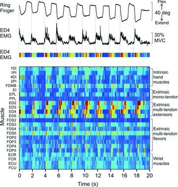
Figure 3. EMG recording from 24 muscle/muscle compartments during unloaded finger movements in a human subject
Subject was instructed to flex and extend the metacarpal phalangeal (MCP) joint of the ring finger paced by a metronome. Top trace shows angular displacement of the MCP joint of the ring finger (digit 4). Second trace from top shows rectified and filtered EMG signal recorded from the digit 4 compartment of extensor digitorum (ED4), one of the prime movers for this task. Third trace shows the ED4 EMG represented as a colour plot with warmer colours indicating higher levels of activity. Each row in the bottom panel shows the activity of one of the 24 muscles recorded over the 20 s of this trial involving 10 repeated extension flexion movements. All EMG signals were normalized to maximum voluntary contraction (MVC). The colour of each small bin within a row indicates the average normalized EMG over a 30 ms epoch (darkest blue = 0%, deep red = ∼30% MVC EMG). EMG signals were recorded from five intrinsic hand muscles (1DI – first dorsal interosseus, 1PI – first palmar interosseus, 4DI – fourth dorsal interosseous, 2PI – second palmar interosseus, FDMB – flexor digiti minimi brevis), three extrinsic hand muscle with a single tendon (EI – extensor indicis, EPL – extensor pollicis longus, FPL – flexor pollicis longus), four compartments of the multitendoned extensor digitorum (ED2–ED5), four compartments each of the multitendoned flexor muscles flexor digitorum superficialis (FDS2–5) and flexor digitorum profundus (FDP2–5), and four wrist muscles (ECR – extensor carpi radialis, FCR – flexor carpi radialis, ECU – extensor carpi ulnaris, FCU – flexor carpi ulnaris). All muscles except FDI and FDMB were recorded with intramuscular electrodes. Many muscles were involved in these unloaded movements of a single joint of an individual finger.
Figure 4
Figure 4. Representative cross-correlation histograms of firing times for pairs of human motor units recorded during the precision grip
A, both units of the pair recorded within the extrinsic thumb muscle, flexor pollicis longus (Thumb–Thumb) of the dominant hand.B, one unit of pair recorded from the thumb muscle and other unit recorded from index finger compartment of flexor digitorum profundus (Thumb–Index) of the dominant hand.C, one unit recorded from the thumb muscle and other unit recorded from index finger muscle (Thumb–Index) of the non-dominant hand. Overall, synchrony for pairs of motor units residing in separate muscles (Thumb–Index) of the dominant hand was just as large as that for pairs of units both within the thumb muscle. The high level of synchrony seen across muscles in the dominant hand, however, was absent in the non-dominant hand. Redrawn from Hockensmith et al. (2005) (with permission of the Society for Neuroscience).
Figure 5
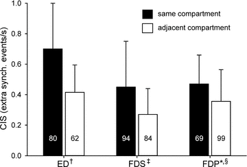
Figure 5. Mean (SD) common input strength (CIS – index representing magnitude of short-term synchrony; Nordstrom et al. 1992) for pairs of motor units residing in the same compartment or adjacent compartments of three human multi-tendoned hand muscles, extensor digitorum (ED), flexor digitorum superficialis (FDS) and flexor digitorum profundus (FDP)
Mean (SD) CIS values: ED same = 0.70 (0.30), ED adjacent = 0.41 (0.18), FDS same = 0.45 (0.30), FDS adjacent = 0.27 (0.17), FDP same = 0.47 (0.19), FDP adjacent = 0.36 (0.21). Values inside of bars indicate number of motor unit pairs. Data compiled from: †Keen & Fuglevand (2004b); ‡McIsaac & Fuglevand (2007); *McIsaac & Fuglevand (unpublished data); §Winges & Santello (2004).

Similar articles

Cited by

References

    1. An KN, Hui FC, Morrey BF, Linscheid RL, Chao EY. Muscles across the elbow joint: a biomechanical analysis. J Biomech. 1981;14:659–669. - PubMed
    1. Bakels R, Kernell D. Average but not continuous speed match between motoneurons and muscle units of rat tibialis anterior. J Neurophysiol. 1993;70:1300–1306. - PubMed
    1. Baker SN. The primate reticulospinal tract, hand function and functional recovery. J Physiol. 2011;589:5603–5612. - PMC - PubMed
    1. Bigland-Ritchie B, Fuglevand AJ, Thomas CK. Contractile properties of human motor units: is man a cat? Neuroscientist. 1998;4:240–249.
    1. Bromberg MB. Updating motor unit number estimation (MUNE) Clin Neurophysiol. 2007;118:1–8. - PubMed

Publication types

LinkOut - more resources