. 2011 Dec 15;589(Pt 24):6105-18.
doi: 10.1113/jphysiol.2011.217497. Epub 2011 Oct 17.

Analysis of pacemaker activity in the human stomach

Affiliations

Analysis of pacemaker activity in the human stomach

Poong-Lyul Rhee et al. J Physiol. .

Abstract

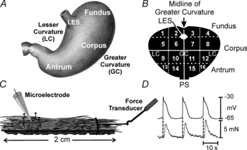
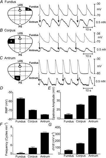
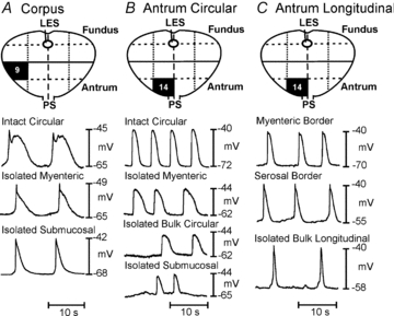
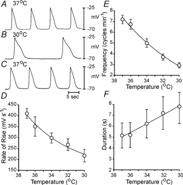
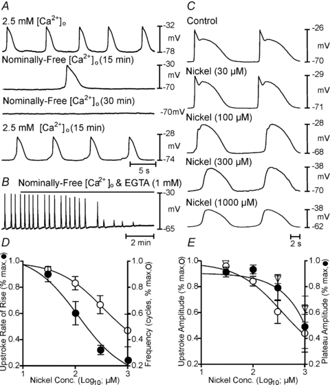
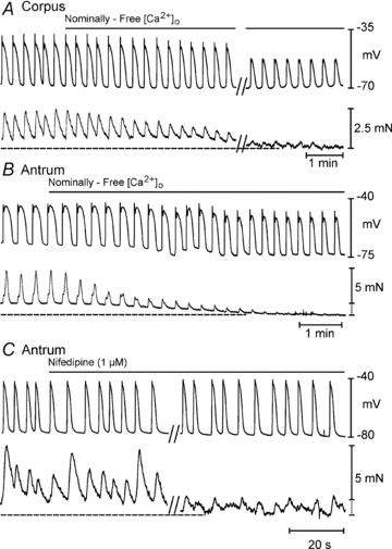
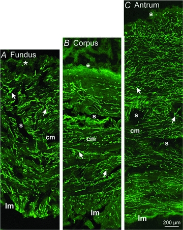
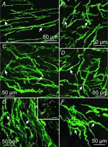
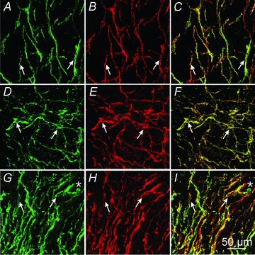
Extracellular electrical recording and studies using animal models have helped establish important concepts of human gastric physiology. Accepted standards include electrical quiescence in the fundus, 3 cycles per minute (cpm) pacemaker activity in corpus and antrum, and a proximal-to-distal slow wave frequency gradient. We investigated slow wave pacemaker activity, contractions and distribution of interstitial cells of Cajal (ICC) in human gastric muscles. Muscles were obtained from patients undergoing gastric resection for cancer, and the anatomical locations of each specimen were mapped by the operating surgeon to 16 standardized regions of the stomach. Electrical slow waves were recorded with intracellular microelectrodes and contractions were recorded by isometric force techniques. Slow waves were routinely recorded from gastric fundus muscles. These events had similar waveforms as slow waves in more distal regions and were coupled to phasic contractions. Gastric slow wave frequency was significantly greater than 3 cpm in all regions of the stomach. Antral slow wave frequency often exceeded the highest frequency of pacemaker activity in the corpus. Chronotropic mechanisms such as muscarinic and prostaglandin receptor binding, stretch, extracelluar Ca(2+) and temperature were unable to explain the observed slow wave frequency that exceeded accepted normal levels. Muscles from all regions through the thickness of the muscularis demonstrated intrinsic pacemaker activity, and this corresponded with the widespread distribution in ICC we mapped throughout the tunica muscularis. Our findings suggest that extracellular electrical recording has underestimated human slow wave frequency and mechanisms of human gastric function may differ from standard laboratory animal models.


Figures

Figure 1
Figure 1. Map of stomach and techniques to record electrical and contractile activity in human stomach
A, sketch of stomach and major regions referred to in text. B, schematic map of stomach used by surgeons to demarcate region from which surgical samples were obtained. Regions 1–4 correspond to the fundus, regions 5–12 correspond to the corpus, and regions 13–16 correspond to the antrum. PS, pyloric sphincter; LES, lower esophageal sphincter. GC and LC represent greater and lesser curvatures, respectively. C, sketch of cross-sectional muscle strip with a microelectrode to record intracellular electrical activity and attachment of a force transducer at one end to record isometric contractions or stretch. D, simultaneous recording from antrum (region 14) showing one-to-one relationship between electrical slow waves and phasic contractions.
Figure 2
Figure 2. Dissections of gastric muscles to obtain subsections for intracellular electrical recordings
A, a phase-contrast image of a montage through the full-thickness of the human gastric antrum muscularis. Note the marked division of the circular muscle layer into bundles separated by broad septae (arrows). Higher magnifications are denoted by arrows. B, full thickness strips cut parallel to either the circular or the longitudinal muscle layers. Strips parallel to the circular muscle were used to cut subregional muscle bundles (C) from the submucosal circular, which consisted of: circular muscle adjacent to the submucosa (a), interior/bulk circular muscle bundles consisting of circular muscle fibres in the central 1/3 of the circular layer (b), or myenteric regions of the circular muscle layer (c). Longitudinal strips were used to cut subregions of longitudinal muscles which were dissected into an inner longitudinal layer with myenteric plexus attached (d) and an outer bulk longitudinal layer adjacent to the serosal surface (e).
Figure 3
Figure 3. Electrical and mechanical activities recorded from various regions of the human stomach
Regions from which specimens were obtained are denoted in surgical maps at left of each panel. A, recording from fundus where slow waves were routinely recorded. Each slow wave was associated with a phasic contraction (arrows) which summed to create tone. B, slow waves and corresponding phasic contractions in corpus muscle. C, slow waves and corresponding phasic contractions from an antral muscle. D–G, summary of electrical parameters (RMP, upstroke amplitude, frequency and rate of rise of the slow wave upstroke) from fundus, corpus and antrum.
Figure 4
Figure 4. Electrical slow waves in subregions of gastric muscle denoted on surgical maps in each panel
A–C, recordings from intact circular (A and B) or longtitudinal (C) muscle strips from near the myenteric border of the circular muscle layer (isolated myenteric), near the submucosal surface of the circular layer (isolated submucosal), the interior region of circular or longitudinal muscles (bulk circular), or longitudinal muscles from near the myenteric border (myenteric longitudinal) or the serosal half of the longitudinal muscle (bulk longitudinal). Slow waves were recorded from all subregions of muscles.
Figure 5
Figure 5. Effects of temperature on slow wave parameters
A–C, slow waves recorded during the same impalement as temperature was decreased from 37°C to 30°C and then returned to 37°C. D–F summarize the effects of temperature on the rate-of-rise of slow waves (D), frequency (E) and duration (F) (5 antral muscles of 3 patients). Data in D–F are fit with exponential functions (D and E, R2 = 0.9990; F, R2 = 0.9994).
Figure 6
Figure 6. Effects of reduced Ca2+ and Ni2+ on slow wave parameters
A, the time-dependent effects during a single impalement of reducing extracellular Ca2+ ([Ca2+]o) from 2.5 mm (control) to nominally free [Ca2+]o (15 and 30 min), and return to 2.5 mm[Ca2+]o. B, effects of adding EGTA (1 mm) to nominally free [Ca2+]o. Slow waves were rapidly blocked when EGTA was added. C, the effects of Ni2+ (0–1000 μm) on slow waves. D and E show summarized effects of Ni2+ on membrane potential (open diamonds in E), slow wave rate-of-rise (IC50 = 114 μm; R2 = 0.99), frequency (IC50 = 339 μm; R2 = 1), upstroke (IC50 = 372 μm; R2 = 0.99) and plateau amplitude (IC50 = 96186 μm; R2 = 0.99).
Figure 7
Figure 7. Effects of reduced [Ca2+]o and nifedipine on slow waves and contractions
A and B, simultaneous recordings of slow waves and phasic contractions in corpus and antrum in response to reduction in [Ca2+]o from 2.5 mm to nominally free [Ca2+]o. Note nearly total inhibition of contractions and slight reduction in amplitude and durations of slow waves. Tone is also reduced in both muscles with the reduction in amplitude. Break in A represents 20 min gap in the recording during a continous impalement. C, response of an antral muscle to nifedipine (1 μm). Note reduction, but not block, of contractions with this compound and little or no effect on slow waves. Break represents 10 min gap in recording during a continuous impalement.
Figure 8
Figure 8. Montages of immunohistochemical confocal images taken through the fundus, corpus and antrum revealing Kit-immunopositive cells
Higher resolution images (see Fig. 9 and Supplemental Figs 4–6) show that the majority of these cells are ICC, but mast cells were also identified by Kit labelling and their rounded appearance (particularly at the submucosal (s) surface of the circular muscle). A–C, a montage through the fundus region (A), the corpus region (B) and the antrum (C). Note the extensive distributions of ICC (arrows) throughout all regions of the circular (cm) and longitudial (lm) muscle bundles that were separated by septae (s). Mast cells (*) are also observed along the submucosal surface of the circular muscle layer.
Figure 9
Figure 9. Higher power confocal images of Kit-positive ICC in different locations through the human stomach
A and B, spindle shaped ICC and ICC with several projections within the gastric fundus (arrows; region 4). C and D, Kit-positive ICC within the gastric corpus (arrows; region 5). ICC formed dense networks throughout this region. E and F, Kit-positive ICC in the gastric antrum (region 13). Dense clusters of spindle shaped ICC and ICC with several projections were observed in the circular layer of this gastric region. Large numbers of small rounded Kit-positive mast cells were observed along the submucosal surface of the human stomach (*, inset in E). Scale bars = 50 μm.
Figure 10
Figure 10. Double labelling of Kit and Ano-1/TMEM16A in the human stomach
A and C, Kit (A, green), Ano-1 (B, red) and merged image (C) of ICC in the gastric fundus (arrows). DF, Kit (D, green), Ano-1 (E, red) and merged image (F) of ICC in the gastric corpus (arrows). G–I, Kit (G, green), Ano-1 (H, red) and merged image (I) in the gastric antrum (arrows). Scale bar in I = 50 μm and represents all panels.

Comment in

  • The analysis of human gastric pacemaker activity.
    O'Grady G, Pullan AJ, Cheng LK. O'Grady G, et al. J Physiol. 2012 Mar 1;590(5):1299-300; author reply 1301-2. doi: 10.1113/jphysiol.2011.224014. J Physiol. 2012. PMID: 22399822 Free PMC article. No abstract available.

Similar articles

Cited by

References

    1. Bayguinov O, Hennig GW, Sanders KM. Movement based artifacts in extracellular electrical recording from GI muscles. Neurogastroenterol Motil. 2011 (in press) - PMC - PubMed
    1. Burns AJ, Lomax AE, Torihashi S, Sanders KM, Ward SM. Interstitial cells of Cajal mediate inhibitory neurotransmission in the stomach. Proc Natl Acad Sci U S A. 1996;93:12008–12013. - PMC - PubMed
    1. Carlson HC, Code CF, Nelson RA. Motor action of the canine gastroduodenal function: a cineradiographic, pressure and electric study. Am J Dig Dis. 1966;11:155–172. - PubMed
    1. Chen JDZ, Lin Z, Pan J, McCallum RW. Abnormal gastric myoelectrical activity and delayed gastric emptying in patients with symptoms suggestive of gastroparesis. Dig Dis Sci. 1996;41:1538–1545. - PubMed
    1. Cousins HM, Edwards FR, Hickey H, Hill CE, Hirst GD. Electrical coupling between the myenteric interstitial cells of Cajal and adjacent muscle layers in the guinea-pig gastric antrum. J Physiol. 2003;550:829–844. - PMC - PubMed

Publication types

LinkOut - more resources