. 2012 May;39(5):1162-70.
doi: 10.1183/09031936.00093911. Epub 2011 Oct 17.

Resolution of acute lung injury and inflammation: a translational mouse model

Affiliations

Resolution of acute lung injury and inflammation: a translational mouse model

Brijesh V Patel et al. Eur Respir J. 2012 May.

Abstract

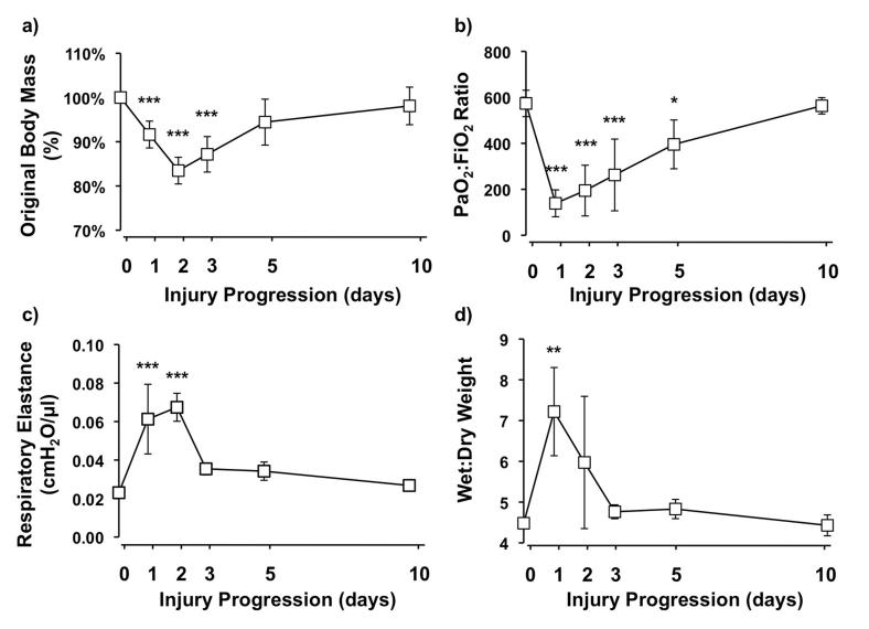
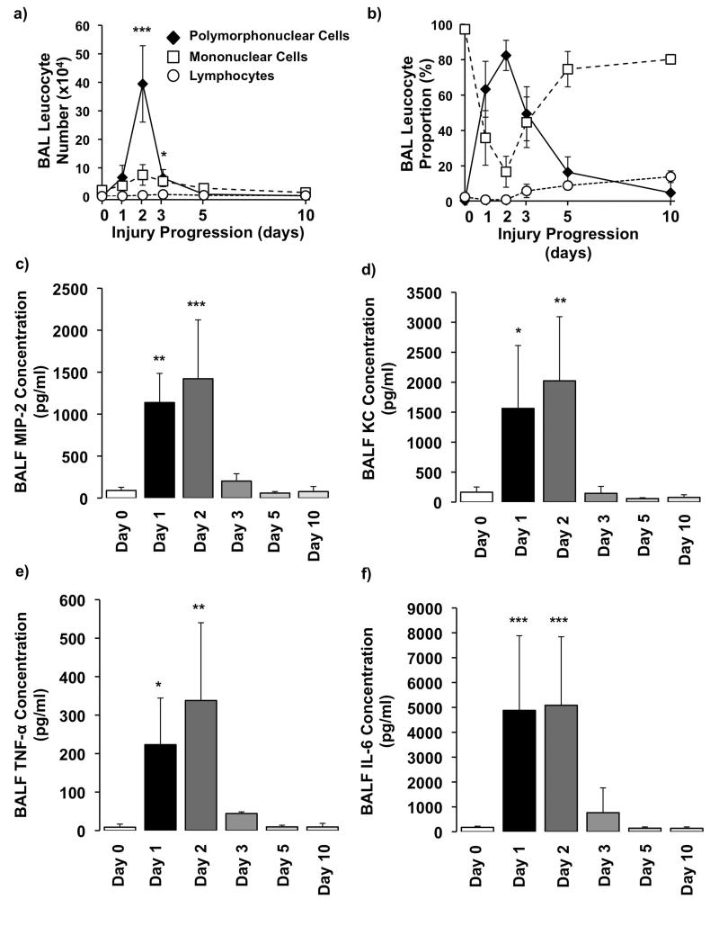
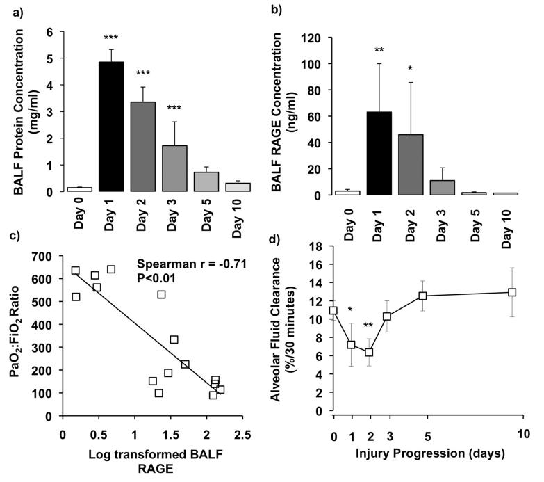
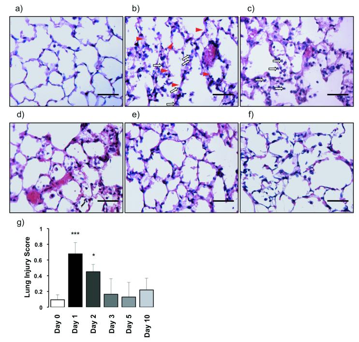
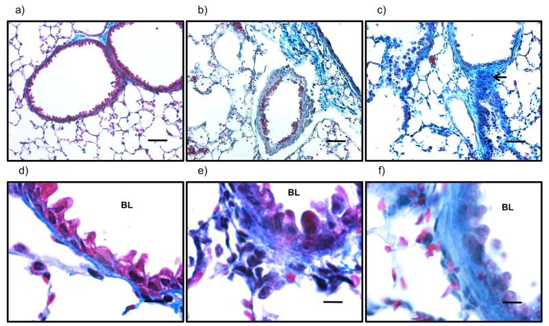
Previous animal models of acute lung injury (ALI) are limited as they only reproduce part of the complex pathobiology of clinical ALI. Here we develop a translational mouse model of ALI, which not only reflects the major clinical and pathological features but also enables investigation into ALI resolution. Anaesthetised mice underwent orotracheal instillation of hydrochloric acid. During the immediate period after instillation, mice were carefully maintained with supplemental oxygen to avoid mortality. At specified time-points, lung injury was assessed by analysis of blood gases, respiratory mechanics, bronchoalveolar lavage fluid, alveolar fluid clearance and lung histology. Animals exhibited significant weight loss, decreased oxygenation, increased respiratory elastance and pulmonary inflammation (intra-alveolar leukocyte influx/cytokine levels and histological injury scores). Moreover, mice displayed alveolar-capillary barrier dysfunction/epithelial injury as reflected by increased alveolar protein, lung wet/dry weight ratio and soluble receptor for advanced glycation end-products, as well as reduced alveolar fluid clearance. These injury parameters peaked between days 1 and 3, followed by almost complete recovery over days 5-10. Histology showed evidence of fibrosis on day 10. The results indicate that this resolving model of acid aspiration represents a powerful experimental tool to investigate the injurious, inflammatory, fibrotic, and resolving and reparative processes of ALI.


Figures

Figure 1
Figure 1
a) Body weight changes as represented by percentage of original body mass (N=9-11/each time point). b) Arterial oxygenation as measured by PaO2:FiO2 at different stages of lung injury (N=5-7/each time point). c) Respiratory system elastance (N=3-5/each time point) and d) lung wet/dry weight ratio (N=3-4/each time point) at different time points after acid instillation. ***P<0.001, ** P<0.01, *P<0.05 vs Day 0 uninjured controls.
Figure 2
Figure 2
BAL a) numbers and b) proportions of polymorphonuclear neutrophils, mononuclear cells, and lymphocytes within the model (N=3-5/each time point).Measurement of BAL fluid (BALF) levels of chemokines c) macrophage inflammatory protein (MIP)-2 and d) keratinocyte-derived chemokine (KC). BALF cytokines e) tumour necrosis factor (TNF)-alpha and f) interleukin (IL)-6 after acid aspiration (N=3-6/each time point). ***P<0.001, ** P<0.01, *P<0.05 vs Day 0 uninjured controls.
Figure 3
Figure 3
a) Changes in alveolar-capillary barrier permeability as measured by BALF protein level (N=3-6/each time point). b) Measurement of BALF soluble RAGE levels as a marker of type 1 alveolar epithelial injury (N=3-6/each time point). c) PaO2:FiO2 plotted against log transformed RAGE shows that oxygenation inversely correlated with the level of alveolar RAGE (R=−0.71, P<0.01). Each data point represents a separate animal. d) Measurement of alveolar fluid clearance rate as a marker of epithelial function (N=3-6/each time point). ***P<0.001, ** P<0.01, * P<0.05 vs Day 0 uninjured controls.
Figure 4
Figure 4
a–f) Representative haematoxylin and eosin (H&E)-stained sections at 6400 original magnification of uninjured and injured animals at all time-points after acid aspiration. a) Day 0, b) day 1, c) day 2, d) day 3, e) day 5 and f) day 10. There is greater cellularity consisting mainly of neutrophils (white arrows) on days 1 and 2 with more areas of atelectasis as well as increased alveolar disruption with hyaline membranes (arrowheads), proteinaceous debris and haemorrhage. With increasing time there is a reduction in cellularity as well as a shift from a neutrophilic to a predominantly mononuclear infiltrate. Scale bars=50μm. g) Lung injury scoring shows a significant injury on days 1 and 2 with a reduction from day 3 onwards (N=3-4/each time point). Lung injury was assessed on a scale of 0–2 for each of the following criteria: i) neutrophils in the alveolar space, ii) neutrophils in the interstitial space, iii) number of hyaline membranes, iv) amount of proteinaceous debris, and v) extent of alveolar septal thickening. The final injury score was derived from the following calculation: Score = [20*(i) + 14*(ii) + 7*(iii) + 7*(iv) + 2*(v)] / (number of fields *100). ***P<0.001, *P<0.05 vs Day 0 uninjured controls.
Figure 5
Figure 5
Representative Masson’s trichrome stained sections at a–c) x200 (scale bars=50μm) and d–f) x1000 (scale bars=10μm) original magnification of uninjured (a and d), and injured animals at days 2 (b and e) and 10 (c and f) after acid aspiration. There are significant changes, with cellular infiltration focussed in the peribronchiolar regions. Higher magnification images show increasing thickness of bronchiolar walls as a result of increased collagen deposition already at day 2 (e), and increasing over the course of the injury. This is particularly apparent at day 10 (c) which shows multiple fibrotic foci (black arrow). BL: bronchiolar lumen.

Comment in

References

    1. Rubenfeld GD, Caldwell E, Peabody E, Weaver J, Martin DP, Neff M, et al. Incidence and outcomes of acute lung injury. N Engl J Med. 2005 Oct 20;353(16):1685–93. - PubMed
    1. Ware LB, Matthay MA. The acute respiratory distress syndrome. N Engl J Med. 2000 May 4;342(18):1334–49. - PubMed
    1. Matute-Bello G, Downey G, Moore BB, Groshong SD, Matthay MA, Slutsky AS, et al. An Official American Thoracic Society Workshop Report: Features and Measurements of Experimental Acute Lung Injury in Animals. Am J Respir Cell Mol Biol. 2011 May;44(5):725–38. 2011. - PMC - PubMed
    1. Matute-Bello G, Frevert CW, Martin TR. Animal models of acute lung injury. Am J Physiol Lung Cell Mol Physiol. 2008 Sep 1;295(3):L379–99. - PMC - PubMed
    1. Ware LB. Modeling human lung disease in animals. Am J Physiol Lung Cell Mol Physiol. 2008 Feb;294(2):L149–50. - PubMed

Publication types

MeSH terms