. 2011 Dec;31(24):4887-901.
doi: 10.1128/MCB.05690-11. Epub 2011 Oct 17.

β-arrestin2 plays permissive roles in the inhibitory activities of RGS9-2 on G protein-coupled receptors by maintaining RGS9-2 in the open conformation

Affiliations

β-arrestin2 plays permissive roles in the inhibitory activities of RGS9-2 on G protein-coupled receptors by maintaining RGS9-2 in the open conformation

Mei Zheng et al. Mol Cell Biol. 2011 Dec.

Abstract

Together with G protein-coupled receptor (GPCR) kinases (GRKs) and β-arrestins, RGS proteins are the major family of molecules that control the signaling of GPCRs. The expression pattern of one of these RGS family members, RGS9-2, coincides with that of the dopamine D(3) receptor (D(3)R) in the brain, and in vivo studies have shown that RGS9-2 regulates the signaling of D2-like receptors. In this study, β-arrestin2 was found to be required for scaffolding of the intricate interactions among the dishevelled-EGL10-pleckstrin (DEP) domain of RGS9-2, Gβ5, R7-binding protein (R7BP), and D(3)R. The DEP domain of RGS9-2, under the permission of β-arrestin2, inhibited the signaling of D(3)R in collaboration with Gβ5. β-Arrestin2 competed with R7BP and Gβ5 so that RGS9-2 is placed in the cytosolic region in an open conformation which is able to inhibit the signaling of GPCRs. The affinity of the receptor protein for β-arrestin2 was a critical factor that determined the selectivity of RGS9-2 for the receptor it regulates. These results show that β-arrestins function not only as mediators of receptor-G protein uncoupling and initiators of receptor endocytosis but also as scaffolding proteins that control and coordinate the inhibitory effects of RGS proteins on the signaling of certain GPCRs.


Figures
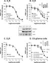
Fig. 1.
Fig. 1.
Effects of RGS9-2 on the signaling of D2-like receptors. (A) Effects of RGS9-2 on the signaling of D2R were determined in cells expressing D2R together with EGFP-Gβ5 (0.6 μg) and/or RGS9-2-EGFP (3 μg). Cellular cAMP was measured using the CRE-Luci (luciferase) reporter gene as described in Materials and Methods. The receptor expression level was maintained at around 1.2 pmol/mg protein. (B) Effects of RGS9-2 on the signaling of D3R were determined in cells expressing D3R as in panel A. The receptor expression level was maintained at around 1.5 pmol/mg protein. #, P < 0.05 for the RGS9-2 group versus the mock-treated group. ###, P < 0.001 for the RGS9-2+Gβ5 group versus the mock-treated group. (C) Effects of RGS9-2 on the signaling of D4R were determined in cells expressing D4R together with EGFP-Gβ5 and RGS9-2-EGFP. The receptor expression level was maintained at around 1.2 pmol/mg protein. Levels of RGS9-2 and Gβ5 expression were determined in the cells used in panels A to C. Cell lysates from each experimental group were separated by SDS-PAGE and blotted with antibodies to GFP and actin. (D) Effects of RGS9-2 on the signaling of D3R and D2R were determined in brain-derived C6 glioma cells expressing D3R or D2R together with Gβ5 and RGS9-2 as described for panels A and B. Receptor expression levels were around 0.7 pmol/mg protein. #, P < 0.05 for the D3R+RGS9-2+Gβ5 group versus the D3R+Mock group. Emax, maximum effect.
Fig. 2.
Fig. 2.
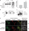
Dopamine D2R and D3R differently interact with RGS9-2. (A) Interaction between RGS9-2 and D2R or D3R was determined by immunoprecipitation. HEK293 cells were transfected with RGS9-2-EGFP together with FLAG-tagged D2R or D3R in pCMV5. Cell lysates were immunoprecipitated (IP) with FLAG beads and immunoblotted (IB) with antibodies to GFP. The receptor expression level was maintained at around 1.2 pmol/mg protein. Receptor proteins were immunoprecipitated with mouse monoclonal antibody FLAG beads, and the resulting immunoprecipitates were blotted with antibodies to rabbit FLAG antibodies. Both D2R and D3R are known to be resolved into 2 or 3 bands by SDS-PAGE (24). The data represent results of three independent experiments with similar outcomes. (B) Determination of interaction between RGS9-2 and D2R or D3R by GST pulldown assay. Bacterial lysates containing the GST fusion proteins of the third intracellular loop of D2R (I3D2-N, the N-terminal part; I3D2-C, the C-terminal part) or D3R (I3D3) were mixed with lysates of HEK293 cells transfected with RGS9-2-EGFP. After three washes, GST beads were incubated with SDS sample buffer. The eluents were analyzed by SDS-PAGE and blotted with antibodies to GFP (GST pulldown part). A blot of HEK293 cell lysates is shown in the lysate part. On the right is an SDS-PAGE analysis of the afterwash of bacterial cell lysates. (C) Colocalization of RGS9-2 and D2R or D3R was determined by immunocytochemistry. Cells were transfected with FLAG-D2R or D3R along with RGS9-2-EGFP (left two panels). Cells were labeled with antibodies to FLAG, followed by Alexa Fluor 594-conjugated anti-rabbit secondary antibodies. In the right panel, cells stably expressing D2R (1.2 pmol/mg protein) or D3R (0.9 pmol/mg protein) were transfected with RGS9-2-EGFP.
Fig. 3.
Fig. 3.
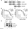
Roles of specific subdomains of RGS9-2 in the regulation of D3R signaling. (A) Schematic diagram of the RGS9-2 constructs (28). The numbers at the top of the diagram indicate the positions of the domains in the original wild-type protein starting from the first Arg residue. (B and C) Role of the DEP domain in the regulation of D3R signaling by RGS9-2. (B) Cells were transfected with 3 μg of the FL-RGS9-2-EGFP or DEPless-EGFP construct together with Gβ5 in pCMV5. Receptor expression levels were equalized for each experimental group (around 1.7 pmol/mg protein). ##, P < 0.01 for the Gβ5+FL group versus the Gβ5+Mock group. (C) Cells were transfected with 3 μg FL-RGS9-2-EGFP and DEP-EGFP. Receptor expression levels were adjusted to around 2.0 pmol/mg protein. #, P < 0.05 for the FL or DEP group versus the mock-treated group. (D) Interactions between the D3R and RGS9-2 proteins were determined by immunoprecipitation from cell lysates expressing FLAG-D3R along with FL-RGS9-2-EGFP, DEPless-RGS9-2-EGFP, and the DEP domain of RGS9-2-EGFP. Immunoprecipitation (IP) and immunoblotting (IB) were conducted with FLAG beads and antibodies to GFP, respectively. Receptor expression levels were adjusted to around 1.7 pmol/mg protein. The data represent results of three independent experiments with similar outcomes. Emax, maximum effect.
Fig. 4.
Fig. 4.
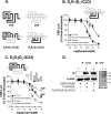
Determination of receptor regions responsible for RGS9-2-mediated inhibition of D3R signaling. (A) Schematic representation of chimeric receptors consisting of D2R and D3R, whose second and third intracellular loops were switched. Signaling of the chimeric receptors consisting of D2R and D3R was compared with that of the corresponding wild-type (WT) receptor: D2R versus D2R-(D3-IC23) (B) or D3R versus D3R-(D2-IC23) (C) Receptor expression levels were adjusted to around 1.5 to 1.7 pmol/mg protein. ###, P < 0.001 for the WT-D3R+RGS9-2+Gβ5 group versus the WT-D3R group or for the Chimera+RGS9-2+Gβ5 group versus the Chimera group. (D) Profiles of RGS9-2 interactions with chimeric receptors were determined by immunoprecipitation (IP) with FLAG beads and immunoblotting (IB) with antibodies to GFP or FLAG. Receptor expression levels were adjusted to around 2.1 pmol/mg protein. Receptor proteins were immunoprecipitated and immunoblotted as described in the legend to Fig. 2A. The data represent results of three independent experiments with similar outcomes. Emax, maximum effect.
Fig. 5.
Fig. 5.
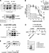
Roles of β-arrestins in the regulatory effects of RGS9-2 on the signaling of D3R. (A) Interactions between β-arrestin2 and RGS9-2 proteins were assessed by immunoprecipitation in cells expressing FLAG-β-arrestin2 along with FL-RGS9-2-EGFP, DEPless-RGS9-2-EGFP, and the DEP domain of RGS9-2-EGFP. Cell lysates were immunoprecipitated (IP) and immunoblotted (IB) with FLAG beads and antibodies to GFP or FLAG, respectively (upper panel). In the lower panel, cells were transfected with RGS9-2-EGFP along with FLAG-β-arrestin1 or FLAG-β-arrestin2. Cell lysates were immunoprecipitated with FLAG beads, and the immunoprecipitates were immunoblotted with GFP or FLAG. The data represent results of three independent experiments with similar outcomes. (B and C) Roles of β-arrestin2 in the regulatory activity of RGS9-2 were determined in β-arrestin2 KD cells. (B) Role of β-arrestin2 in the inhibitory effect of RGS9-2 on the signaling of D3R. Receptor expression levels were maintained at around 1.8 to 2.0 pmol/mg protein. ##, P < 0.01 for the Con-KD+RGS9-2/Gβ5 group versus the Con-KD+Mock or β-arr2-KD+Mock group. Expression of endogenous β-arrestin2 was inhibited through β-arrestin2 shRNA expression in plasmid pcDNA3.0(Neo) (data not shown). Cell lysates were immunoblotted with antibodies to GFP (RGS9-2 and Gβ5), β-arrestin2, and actin. Con, control. (C) Role of β-arrestin2 in the interaction between the D3R and RGS9-2 proteins. Immunoprecipitations from β-arr2-KD cells expressing FLAG-D3R along with FL-RGS9-2-EGFP, DEPless RGS9-2-EGFP, and the DEP domain of RGS9-2-EGFP were conducted. Cell lysates were immunoprecipitated and immunoblotted with FLAG beads and antibodies to GFP, respectively. Receptor expression levels were maintained at around 1.9 pmol/mg protein. The data represent results of three independent experiments with similar outcomes. (D) The role of β-arrestin2 in the interaction between RGS9-2 and D3R was determined with a GST pulldown assay. Bacterial lysates containing the GST fusion proteins of the third intracellular loops of D3R (I3D3) were mixed with lysates of Con-KD or β-arr2-KD cells, which were transfected with RGS9-2-EGFP. After three washes, GST beads were incubated with SDS sample buffer. The eluents were analyzed by SDS-PAGE and blotted with antibodies to GFP (GST pulldown part). A blot of HEK293 cell lysates is shown in the lysate part. (E) Interaction between β-arrestin2 and the DEP domain was determined in a GST pulldown assay. Bacterial lysates containing the GST fusion proteins of the FL protein (GST-β-arr2-FL), the N domain (GST-β-arr2-N), the C domain (GST-β-arr2-C), or the C domain plus the carboxy tail of rat β-arrestin2 (GST-β-arr2-C-CT) were mixed with lysates of HEK293 cells transfected with DEP-EGFP. After three washes, GST beads were incubated with SDS sample buffer. The eluents were analyzed by SDS-PAGE and blotted with antibodies to GFP (GST pulldown part). A blot of HEK293 cell lysates is shown in the lysate part. On the right is an SDS-PAGE analysis of the afterwash of bacterial cell lysates. Emax, maximum effect.
Fig. 6.
Fig. 6.
Role of affinity for β-arrestin2 in the regulation of D3R signaling through RGS9-2. (A) Interaction between β-arrestin2 and D3R was determined by GST pulldown assay. Bacterial lysates containing the GST fusion proteins of the third intracellular loop of D2R or D3R were mixed with lysates of HEK293 cells transfected with β-arrestin2. After three washes, GST beads were incubated with SDS sample buffer. The eluents were analyzed by SDS-PAGE and blotted with antibodies to GFP (GST pulldown part). A blot of HEK293 cell lysates is shown in the lysate part. On the right is an SDS-PAGE analysis of the afterwash of bacterial cell lysates. (B and C) Comparison of the profiles of β-arrestin2 interactions with D2R and D3R in cells expressing β-arrestin2 and FLAG-D2R or FLAG-D3R (B) or with D3R and D3R-(D2-IC23) in cells expressing β-arrestin2 and FLAG-D3R or FLAG-D3R-(D2-IC23) (C). Cells were treated with 10 μM dopamine for 5 min and blotted with antibodies to β-arrestin2. Receptor expression levels were maintained at around 1.5 to 1.7 pmol/mg protein. Receptor proteins were immunoprecipitated (IP) and immunoblotted (IB) as described in the legend to Fig. 2A. The data represent results of three independent experiments with similar outcomes. (D) Role of β-arrestin2 in the regulatory effect of RGS9-2 on the signaling of D3R-(D2-IC23) as determined in β-arrestin2 KD cells in comparison with that in control cells (Fig. 4C). Receptor expression levels were around 1.7 pmol/mg protein. (E) Role of affinity for β-arrestin2 in the regulatory effects of RGS9-2 on the signaling of D2R as determined in cells expressing D2R-β-arr2, a fusion of D2R and β-arrestin2. Receptor expression levels were around 0.6 pmol/mg protein. ###, P < 0.001 for the Gβ5+RGS9-2 group versus the mock-treated group. EC50, 50% effective concentration.
Fig. 7.
Fig. 7.
Role of β-arrestin2 in the interaction of Gβ5 with adjacent proteins. (A) Cooperative activities of β-arrestin2 and RGS9-2 in the inhibition of D3R signaling. Cells were transfected with low doses of β-arrestin2 (1 μg) and RGS9-2 (1 μg). Receptor expression levels were maintained at around 1.9 pmol/mg protein. Cell lysates were immunoblotted with antibodies to GFP, β-arrestin2, and actin. #, P < 0.05 for the β-arr2+RGS9-2/Gβ5 group versus the β-arr2 or RGS9-2/Gβ5 group. (B) Interaction between β-arrestin2 and Gβ5 was determined by immunoprecipitation. Lysates of cells transfected with EGFP-Gβ5 and/or FLAG-β-arrestin2 were immunoprecipitated (IP) with antibodies to FLAG and immunoblotted (IB) with antibodies to GFP. (C) Selective interaction between Gβ5 and D3R. Lysates of cells transfected with EGFP-Gβ5 and FLAG-D2R or FLAG-D3R were immunoprecipitated with antibodies to FLAG and immunoblotted with antibodies to GFP. Receptor proteins were immunoprecipitated and immunoblotted as described in the legend to Fig. 2A. (D) Effects of β-arrestin2 on the interaction between the DEP domain and Gβ5 were determined in cells stably expressing control shRNA or β-arrestin2 shRNA. The cells were additionally transfected with EGFP-Gβ5 and FLAG-DEP. The data represent results of three independent experiments with similar outcomes. (E) Effects of β-arrestin2 on the interaction between D3R and Gβ5 were determined in cells expressing FLAG-D3R and EGFP-Gβ5. Immunoprecipitation and immunoblotting were conducted with FLAG beads and antibodies to GFP, respectively. Receptor expression levels were maintained at around 1.9 pmol/mg protein by [3H]sulpiride binding. Immunoprecipitated receptor proteins were immunoblotted as described in the legend to Fig. 2A. The data represent results of two independent experiments with similar outcomes. (F) Competitive binding of β-arrestin2 by the DEP domain and Gβ5. Cells expressing FLAG-β-arrestin2 and DEP-EGFP were transfected with EGFP-Gβ5. Cell lysates were immunoprecipitated with FLAG beads and blotted with GFP or FLAG antibodies. The data represent results of three independent experiments with similar outcomes. Emax, maximum effect.
Fig. 8.
Fig. 8.
Roles of β-arrestin2 in the subcellular localization of RGS9-2. (A) Effects of KD of endogenous β-arrestins on the subcellular localization of RGS9-2. Con-KD and β-arr2-KD cells were transfected with RGS9-2-EGFP. (B) Effects of R7BP on the subcellular localization of RGS9-2. HEK293 cells were transfected with RGS9-2-EGFP and/or R7BP. (C) Competitive binding of RGS9-2 and R7BP to β-arrestin2. Cells expressing FLAG-β-arrestin2 and RGS9-2-EGFP were transfected with increasing amounts of R7BP-EGFP. Cell lysates were immunoprecipitated (IP) with antibodies to the FLAG epitope and immunoblotted (IB) with antibodies to GFP and FLAG. The data represent results of three independent experiments with similar outcomes. (D) Competitive binding of the DEP domain and R7BP to β-arrestin2. Cells expressing FLAG-β-arrestin2 and DEP-EGFP were transfected with increasing amounts of R7BP-EGFP. Cell lysates were immunoprecipitated with antibodies to the FLAG epitope and immunoblotted with antibodies to GFP and FLAG. (E) Competitive binding of β-arrestin2 and R7BP to the DEP domain of RGS9-2. Cells expressing FLAG-DEP and RGS9-2-EGFP were transfected with increasing amounts of β-arrestin2. Cell lysates were immunoprecipitated with FLAG beads and immunoblotted with antibodies to GFP and FLAG. The data represent results of three independent experiments with similar outcomes. (F) Roles of β-arrestins and R7BP in the regulatory effects of RGS9-2 on the signaling of D3R. HEK293 cells in which β-arrestin1 and β-arrestin2 were simultaneously knocked down (data not shown) were transfected with D3R and different combinations of RGS9-2, Gβ5, β-arrestin2, and R7BP. Cellular cAMP levels were determined as described in the legend to Fig. 1, and the levels of RGS9-2 expression were determined by immunoblotting with antibodies to GFP and actin. ***, P < 0.001 for the Gβ5 group versus the Gβ5+RGS9-2+β-arr2 group; ##, P < 0.01 for the Gβ5+RGS9-2+β-arr2 group versus the Gβ5+RGS9-2 or Gβ5+RGS9-2+β-arr2+R7BP group.
Fig. 9.
Fig. 9.
Proposed working model of the D3R regulatory complex. When Gβ5 and the DEP domain of the RGS9 protein are associated, RGS9-2 is in the inactive (closed) conformation and cannot regulate G protein cycling. If the interaction between Gβ5 and the DEP domain is disrupted, the conformation of the RGS protein is converted to the active (open) conformation (33). RGS9-2 forms a stable complex with Gβ5 via its GGL domain (1). The DHEX linker, but not the DEP domain, directly interacts with Gβ5 (8), and β-arrestin mediates the interaction between the DEP domain and Gβ5 (Fig. 7D). The opposite surface of Gβ5 makes transient/dynamic contacts with the DHEX linker (direct interaction) and the DEP domain (mediated by β-arrestin2) (32) (Fig. 7D and F). This transient contact is disrupted when R7BP interacts with the RGS-Gβ5 complex in the cleft formed between Gβ5 and the DHEX linker region (32). R7BP anchors the RGS9-2/Gβ5 complex on the plasma membrane and stabilizes RGS9-2. R7BP competes with β-arrestin2 for binding with the DEP domain (Fig. 8E) and functionally antagonizes the scaffolding activity of β-arrestin2 for the regulation of D3R signaling (Fig. 8F). Overall, β-arrestin2 converts the RGS9-2/Gβ5 complex to the active conformation by binding to the DEP domain and Gβ5. Also, β-arrestin2 binds with the DEP domain in competition with R7BP, rendering RGS9-2 in the active (open) conformation in the cytosol.

Similar articles

Cited by

References

    1. Accili D., et al. 1996. A targeted mutation of the D3 dopamine receptor gene is associated with hyperactivity in mice. Proc. Natl. Acad. Sci. U. S. A. 93:1945–1949 - PMC - PubMed
    1. Anderson G. R., Posokhova E., Martemyanov K. A. 2009. The R7 RGS protein family: multi-subunit regulators of neuronal G protein signaling. Cell Biochem. Biophys. 54:33–46 - PMC - PubMed
    1. Ballon D. R., et al. 2006. DEP domain-mediated regulation of GPCR signaling responses. Cell 126:1079–1093 - PubMed
    1. Benovic J. L., et al. 1987. Functional desensitization of the isolated beta-adrenergic receptor by the beta-adrenergic receptor kinase: potential role of an analog of the retinal protein arrestin (48-kDa protein). Proc. Natl. Acad. Sci. U. S. A. 84:8879–8882 - PMC - PubMed
    1. Beom S., Cheong D., Torres G., Caron M. G., Kim K. M. 2004. Comparative studies of molecular mechanisms of dopamine D2 and D3 receptors for the activation of extracellular signal-regulated kinase. J. Biol. Chem. 279:28304–28314 - PubMed

Publication types

MeSH terms

LinkOut - more resources