

Anterior cruciate ligament changes in the human knee joint in aging and osteoarthritis
- PMID: 22006159
- PMCID: PMC3266452
- DOI: 10.1002/art.33417
Anterior cruciate ligament changes in the human knee joint in aging and osteoarthritis
Abstract
Objective: The development and patterns of spontaneous age-related changes in the anterior cruciate ligament (ACL) and their relationship to articular cartilage degeneration are not well characterized. This study was undertaken to investigate the types and temporal sequence of age-related ACL changes and to determine their correlation with cartilage lesion patterns at all stages of osteoarthritis (OA) development in human knee joints without prior joint trauma.
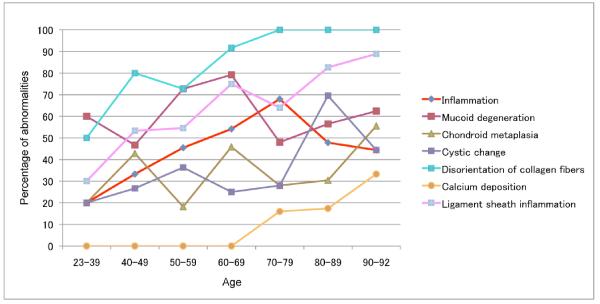
Methods: Human knee joints (n = 120 from 65 donors ages 23-92) were obtained at autopsy, and ACLs and cartilage were graded macroscopically and histologically. Inflammation surrounding the ACL was assessed separately.
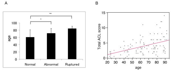
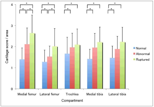
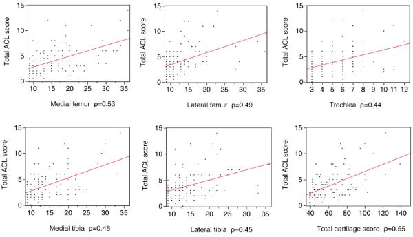
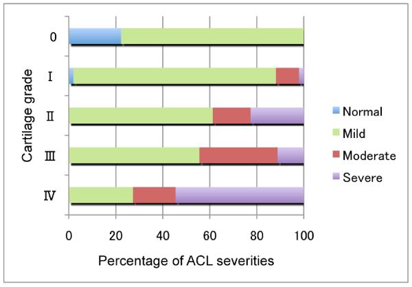
Results: Histologic ACL substance scores and ligament sheath inflammation scores increased with age. Collagen fiber disorganization was the earliest and most prevalent change. The severity of mucoid degeneration and chondroid metaplasia in the ACL increased with the development of cartilage lesions. A correlation between ACL degeneration and cartilage degeneration was observed, especially in the medial compartment of the knee joint.
Conclusion: Our findings indicate that ACL degeneration is highly prevalent in knees with cartilage defects and may even precede cartilage changes. Hence, ACL deficiencies may not only be important in posttraumatic OA, but may also be a feature associated with knee OA pathogenesis in general.
Copyright © 2012 by the American College of Rheumatology.
Figures
References
-
- Felson DT, Lawrence RC, Dieppe PA, Hirsch R, Helmick CG, Jordan JM. Osteoarthritis: new insights. Part 1: the disease and its risk factors. Ann Intern Med. 2000;17(133):635–46. - PubMed
-
- Felson DT, Zhang Y, Hannan MT, Naimark A, Weissman B, Aliabadi P. Risk factors for incident radiographic knee osteoarthritis in the elderly: the Framingham Study. Arthritis Rheumatol. 1997;40:728–33. - PubMed
-
- Hunter DJ, Zhang Y, Niu J, Tu X, Amin S, Goggins J. Structural factors associated with malalignment in knee osteoarthritis: the Boston osteoarthritis knee study. J Rheumatol. 2005;32:2192–9. - PubMed
-
- Cooper C, Snow S, McAlindon TE, Kellingray S, Stuart B, Coggon D. Risk factors for the incidence and progression of radiographic knee osteoarthritis. Arthritis Rheum. 2000;43:995–1000. - PubMed
-
- Bennett LD, Buckland-Wright JC. Meniscal and articular cartilage changes in knee osteoarthritis: a cross-sectional double-contrast macroradiographic study. Rheumatology (Oxford) 2002;41:917–23. - PubMed
Publication types
MeSH terms
Grants and funding
LinkOut - more resources
Full Text Sources
Medical
