Review
. 2012 Feb;14(1):30-8.
doi: 10.1007/s11926-011-0215-5.

Dendritic cells: novel players in fibrosis and scleroderma

Affiliations
Review

Dendritic cells: novel players in fibrosis and scleroderma

Theresa T Lu. Curr Rheumatol Rep. 2012 Feb.

Abstract

Dendritic cells are professional antigen-presenting cells that are most studied for their function in mediating T-cell tolerance and T-cell activation. In addition, recent evidence indicates that dendritic cells can regulate the vasculature and function of fibroblast-type cells. The potential contribution of dendritic cells to scleroderma and fibrosis is not well-understood. In this article, we review recent studies as well as describe our own ongoing work that points toward a role for dendritic cells in scleroderma and fibrosis.


Conflict of interest statement

Disclosure No potential conflicts of interest relevant to this article were reported.

Figures

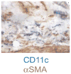
Fig. 1
Fig. 1
Bleomycin-induced skin fibrosis is associated with accumulation of CD11cbright cells with myofibroblasts. Mice received 60 μg of subcutaneous bleomycin in the lower back 5 days per week starting at day 0. Skin was harvested at 28 days and stained for CD11c (blue) and α smooth muscle actin (αSMA) (brown). Note the CD11cbright cells intermingled with the scattered brown αSMA+ myofibroblasts in the deep dermis directly above the panniculus carnosus muscle. Photo was taken at 60× magnification
Fig. 2
Fig. 2
Possible roles of dendritic cells (DCs) in scleroderma and fibrosis. Nonplasmacytoid dendritic cells (blue) could potentially modulate fibrosis by a) expressing proinflammatory cytokines that can activate fibroblastic cells and that contribute to the inflammatory milieu, which may help to drive fibrosis or recovery from fibrosis; b) expressing interleukin (IL)-10 that could potentially contribute to skewing of T-cell responses toward a profibrotic T-helper type 2 (Th2) phenotype and to plasma cell differentiation, but that could also have antifibrotic effects; c) expressing transforming growth factor (TGF)-β that promotes myofibroblast differentiation; and d) modulating vascular growth and function. Plasmacytoid DCs (green) may also contribute via e) expression of type 1 interferons, which can stimulate nonplasmacytoid DCs and plasma cell development. The plasma cells lead to the generation of more autoantibodies, which, in conjunction with Toll-like receptor (TLR) ligands, can be stimulatory for DCs. The thicker lines delineating possibilities a, b, and e indicate those that are supported by recent studies. TNF tumor necrosis factor

References

    1. Varga J, Abraham D. Systemic sclerosis: a prototypic multisystem fibrotic disorder. J Clin Invest. 2007;117:557–67. - PMC - PubMed
    1. Maricq HR, LeRoy EC. Patterns of finger capillary abnormalities in connective tissue disease by “wide-field” microscopy. Arthritis Rheum. 1973;16:619–28. - PubMed
    1. Hettema ME, Zhang D, Stienstra Y, et al. Decreased capillary permeability and capillary density in patients with systemic sclerosis using large-window sodium fluorescein videodensitometry of the ankle. Rheumatology. 2008;47:1409–12. - PubMed
    1. Fleming JN, Nash RA, McLeod DO, et al. Capillary regeneration in scleroderma: stem cell therapy reverses phenotype? PLoS One. 2008;3:e1452. - PMC - PubMed
    1. Ramos-Casals M, Fonollosa-Pla V, Brito-Zeron P, et al. Targeted therapy for systemic sclerosis: how close are we? Nat Rev Rheumatol. 2010;6:269–78. - PubMed