

Active Bax and Bak are functional holins
- PMID: 22006182
- PMCID: PMC3219232
- DOI: 10.1101/gad.171645.111
Active Bax and Bak are functional holins
Abstract
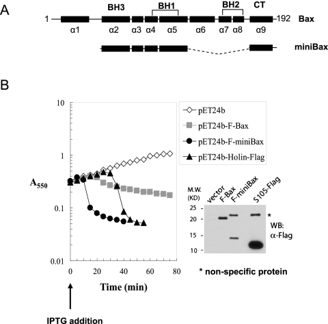
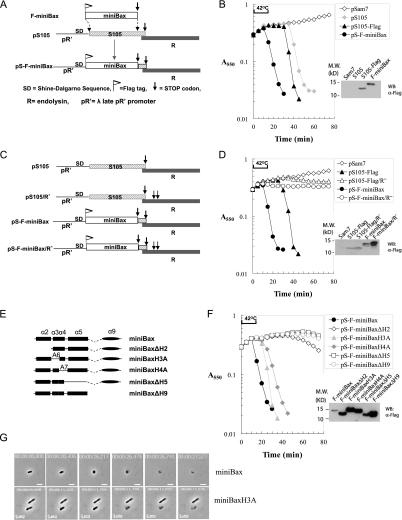
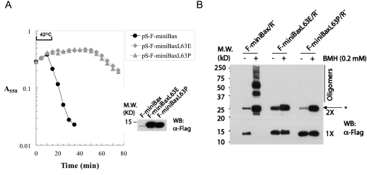
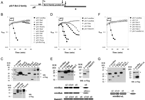
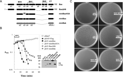
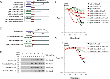
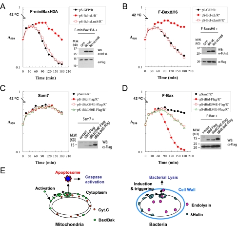
The mechanism of Bax/Bak-dependent mitochondrial outer membrane permeabilization (MOMP), a central apoptotic event primarily controlled by the Bcl-2 family proteins, remains not well understood. Here, we express active Bax/Bak in bacteria, the putative origin of mitochondria, and examine their functional similarities to the λ bacteriophage (λ) holin. As critical effectors for bacterial lysis, holin oligomers form membrane lesions, through which endolysin, a muralytic enzyme, escapes the cytoplasm to attack the cell wall at the end of the infection cycle. We found that active Bax/Bak, but not any other Bcl-2 family protein, displays holin behavior, causing bacterial lysis by releasing endolysin in an oligomerization-dependent manner. Strikingly, replacing the holin gene with active alleles of Bax/Bak results in plaque-forming phages. Furthermore, we provide evidence that active Bax produces large membrane holes, the size of which is controlled by structural elements of Bax. Notably, lysis by active Bax is inhibited by Bcl-xL, and the lysis activity of the wild-type Bax is stimulated by a BH3-only protein. Together, these results mechanistically link MOMP to holin-mediated hole formation in the bacterial plasma membrane.
Figures
Similar articles
-
Inactivation of prosurvival Bcl-2 proteins activates Bax/Bak through the outer mitochondrial membrane.Genes Dev. 2016 Apr 15;30(8):973-88. doi: 10.1101/gad.276725.115. Epub 2016 Apr 7. Genes Dev. 2016. PMID: 27056669 Free PMC article.
-
A Small-Molecule Inhibitor of Bax and Bak Oligomerization Prevents Genotoxic Cell Death and Promotes Neuroprotection.Cell Chem Biol. 2017 Apr 20;24(4):493-506.e5. doi: 10.1016/j.chembiol.2017.03.011. Epub 2017 Apr 6. Cell Chem Biol. 2017. PMID: 28392146 Free PMC article.
-
Regulation of stress-induced nuclear protein redistribution: a new function of Bax and Bak uncoupled from Bcl-x(L).Cell Death Differ. 2010 Feb;17(2):346-59. doi: 10.1038/cdd.2009.145. Epub 2009 Oct 9. Cell Death Differ. 2010. PMID: 19816507
-
Physiological and Pharmacological Control of BAK, BAX, and Beyond.Trends Cell Biol. 2016 Dec;26(12):906-917. doi: 10.1016/j.tcb.2016.07.002. Epub 2016 Aug 4. Trends Cell Biol. 2016. PMID: 27498846 Free PMC article. Review.
-
How do Bax and Bak lead to permeabilization of the outer mitochondrial membrane?Curr Opin Cell Biol. 2006 Dec;18(6):685-9. doi: 10.1016/j.ceb.2006.10.004. Epub 2006 Oct 12. Curr Opin Cell Biol. 2006. PMID: 17046225 Review.
Cited by
-
Use of microfluidic technology to analyze gene expression during Staphylococcus aureus biofilm formation reveals distinct physiological niches.Appl Environ Microbiol. 2013 Jun;79(11):3413-24. doi: 10.1128/AEM.00395-13. Epub 2013 Mar 22. Appl Environ Microbiol. 2013. PMID: 23524683 Free PMC article.
-
Genetically Encoded Fluorescent Biosensor for Rapid Detection of Protein Expression.ACS Synth Biol. 2020 Nov 20;9(11):2955-2963. doi: 10.1021/acssynbio.0c00407. Epub 2020 Oct 12. ACS Synth Biol. 2020. PMID: 33044070 Free PMC article.
-
Bacteriophage-encoded lethal membrane disruptors: Advances in understanding and potential applications.Front Microbiol. 2022 Oct 26;13:1044143. doi: 10.3389/fmicb.2022.1044143. eCollection 2022. Front Microbiol. 2022. PMID: 36345304 Free PMC article. Review.
-
Programmed cell death in bacteria and implications for antibiotic therapy.Trends Microbiol. 2013 Jun;21(6):265-70. doi: 10.1016/j.tim.2013.04.001. Epub 2013 May 14. Trends Microbiol. 2013. PMID: 23684151 Free PMC article.
-
Genome Sequence Comparisons between Small and Large Colony Phenotypes of Equine Clinical Isolates of Arcanobacterium hippocoleae.Animals (Basel). 2024 May 29;14(11):1609. doi: 10.3390/ani14111609. Animals (Basel). 2024. PMID: 38891657 Free PMC article.
References
-
- Agu CA, Klein R, Lengler J, Schilcher F, Gregor W, Peterbauer T, Blasi U, Salmons B, Gunzburg WH, Hohenadl C 2007. Bacteriophage-encoded toxins: the λ-holin protein causes caspase-independent non-apoptotic cell death of eukaryotic cells. Cell Microbiol 9: 1753–1765 - PubMed
-
- Antignani A, Youle RJ 2006. How do Bax and Bak lead to permeabilization of the outer mitochondrial membrane? Curr Opin Cell Biol 18: 685–689 - PubMed
-
- Antonsson B, Montessuit S, Sanchez B, Martinou JC 2001. Bax is present as a high molecular weight oligomer/complex in the mitochondrial membrane of apoptotic cells. J Biol Chem 276: 11615–11623 - PubMed
Publication types
MeSH terms
Substances
Grants and funding
LinkOut - more resources
Full Text Sources
Other Literature Sources
Molecular Biology Databases
Research Materials