. 2011 Oct 25;108(43):17598-603.
doi: 10.1073/pnas.1114746108. Epub 2011 Oct 17.

Compact and flexible raster scanning multiphoton endoscope capable of imaging unstained tissue

Affiliations

Compact and flexible raster scanning multiphoton endoscope capable of imaging unstained tissue

David R Rivera et al. Proc Natl Acad Sci U S A. .

Abstract

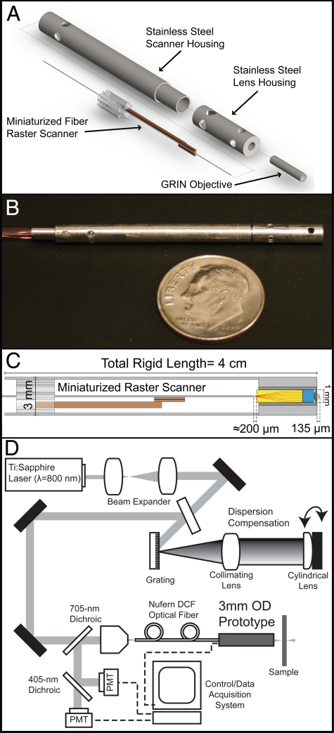
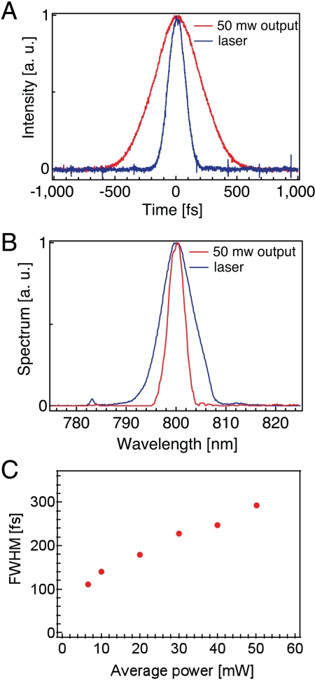
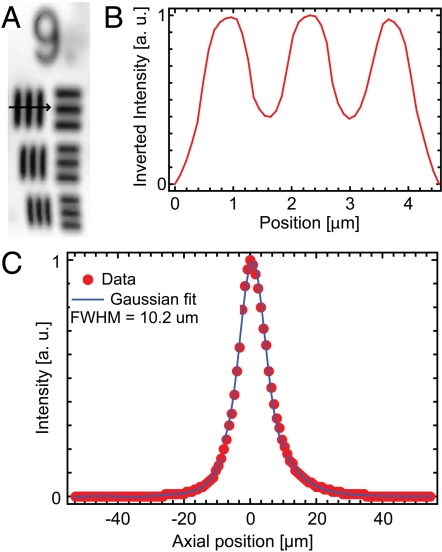
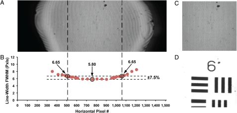
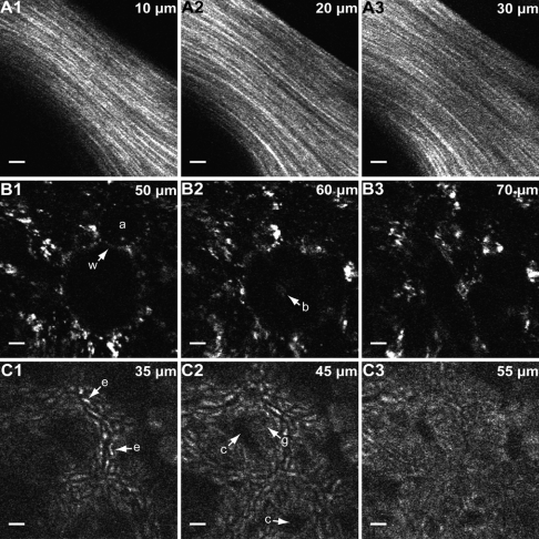
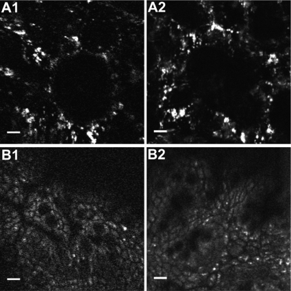
We present a compact and flexible endoscope (3-mm outer diameter, 4-cm rigid length) that utilizes a miniaturized resonant/nonresonant fiber raster scanner and a multielement gradient-index lens assembly for two-photon excited intrinsic fluorescence and second-harmonic generation imaging of biological tissues. The miniaturized raster scanner is fabricated by mounting a commercial double-clad optical fiber (DCF) onto two piezo bimorphs that are aligned such that their bending axes are perpendicular to each other. Fast lateral scanning of the laser illumination at 4.1 frames/s (512 lines per frame) is achieved by simultaneously driving the DCF cantilever at its resonant frequency in one dimension and nonresonantly in the orthogonal axis. The implementation of a DCF into the scanner enables simultaneous delivery of the femtosecond pulsed 800-nm excitation source and epi-collection of the signal. Our device is able to achieve a field-of-view (FOV(xy)) of 110 μm by 110 μm with a highly uniform pixel dwell time. The lateral and axial resolutions for two-photon imaging are 0.8 and 10 μm, respectively. The endoscope's imaging capabilities were demonstrated by imaging ex vivo mouse tissue through the collection of intrinsic fluorescence and second-harmonic signal without the need for staining. The results presented here indicate that our device can be applied in the future to perform minimally invasive in vivo optical biopsies for medical diagnostics.


Conflict of interest statement

The authors declare no conflict of interest.

Figures

Fig. 1.
Fig. 1.
Three-millimeter o.d. raster scanning endoscope components and setup. (A) Mechanical assembly of the endoscope components. (B) Photograph of the prototype. (C) Optical path of the internal components of the distal end. (D) Tabletop endoscope imaging setup.
Fig. 2.
Fig. 2.
Pulse characterization. (A) Autocorrelation pulse-width measurements for the Ti:Sapphire output and for 50-mW endoscope output. The measured pulse widths are approximately 100 and 290 fs, respectively. (B) Spectra for the Ti:Sapphire output and for endoscope output at 50 mW. (C) Measured pulse widths of the endoscope output as a function of output power, assuming a deconvolution factor of 1.54 for a Sech2 pulse.
Fig. 3.
Fig. 3.
Lateral and axial resolution. (A) Transmission image of high-resolution USAF test target. Group 9, elements 1–3 are displayed with line widths ranging from 977–775 nm. FOVxy = 10 μm × 30 μm. (B) Intensity line profile across the vertical bars of group 9, element 1. Using the intensity ratios between the peak and trough values, we determine a one-photon lateral resolution of approximately 1.1 μm (FWHM), which corresponds to a two-photon lateral resolution of approximately 0.8 μm (FWHM). (C) A two-photon axial resolution of approximately 10 μm (FWHM of the thin film response) is determined using a RhB thin film scan.
Fig. 4.
Fig. 4.
Raster scan uniformity characterization. (A) Transmission image of a 400 LP/mm Ronchi ruling (line width = 1.25 μm). (B) Plot of the measured line widths of the Ronchi ruling as a function of the horizontal pixel number across the image. A horizontal range of approximately 110 μm corresponds to a line-width deviation of ± 7.5%. (C) Cropped and postprocessed Ronchi ruling image from Fig. 4A (FOVxy≈110 μm × 110 μm). The Ronchi ruling lines in C are corrected to be of uniform width across the image FOVxy. (D) Cropped and postprocessed transmission image of USAF test target group 6, elements 2–3 (line widths: 7.0 to 6.2 μm, FOVxy≈110 μm × 110 μm).
Fig. 5.
Fig. 5.
TPF/SHG images of ex vivo mouse tissue. (A) Unaveraged SHG images of mouse tail tendon at 10, 20, and 30 μm from the surface. (B) Unaveraged intrinsic fluorescence images of mouse lung at 50, 60, and 70 μm from the tissue surface. In B1, alveolar walls (w) and lumens (a) are clearly visible; in B2, a bronchiole (b) is distinguishable. (C) Five frames averaged intrinsic fluorescence images of mouse colon at 35, 45, and 55 μm from the surface. In C1, enterocytes (e) are visible; in C2, crypts (c) and goblet cells (g) are present. Scale bars, 10 μm. All images were acquired at 4.1 frames/second, 800-nm excitation, and the uniformly sampled FOVxy of approximately 110 μm × 110 μm is displayed. For the images in A, the power at the sample is approximately 30 mW; for the images in B and C, the power at the sample is 75 mW.
Fig. 6.
Fig. 6.
Imaging comparison between the multiphoton endoscope and a commercial multiphoton microscope. (A) Unaveraged intrinsic fluorescence images of ex vivo mouse lung. A1 shows the image acquired from the multiphoton endoscope, and A2 shows the image acquired from the Olympus multiphoton microscope. (B) Five frames averaged intrinsic fluorescence images of ex vivo mouse colon. B1 shows the image acquired from the multiphoton endoscope, and B2 shows the image acquired from the Olympus multiphoton microscope. Scale bars, 10 μm. Comparable amounts of two-photon excited fluorescence at the sample per frame were maintained in each system. The displayed images were acquired with 800-nm excitation and a FOVxy of approximately 110 μm × 110 μm.

Similar articles

Cited by

References

    1. Zipfel WR, et al. Live tissue intrinsic emission microscopy using multiphoton-excited native fluorescence and second harmonic generation. Proc Natl Acad Sci USA. 2003;100:7075–7080. - PMC - PubMed
    1. Mukherjee S, et al. Human bladder cancer diagnosis using Multiphoton microscopy. Proc Soc Photo Opt Instrum Eng. 2009;7161:716117-1–716117-10. - PMC - PubMed
    1. Pavlova I, et al. Multiphoton microscopy as a diagnostic imaging modality for lung cancer. Proc Soc Photo Opt Instrum Eng. 2010;7569:756918-1–756918-7. - PMC - PubMed
    1. Wang CC, et al. Differentiation of normal and cancerous lung tissues by multiphoton imaging. J Biomed Opt. 2009;14:044034-1–044034-4. - PubMed
    1. Wilder-Smith P, et al. In vivo multiphoton fluorescence imaging: A novel approach to oral malignancy. Lasers Surg Med. 2004;35:96–103. - PubMed

Publication types