

Involvement of O-glycosylation defining oncofetal fibronectin in epithelial-mesenchymal transition process
- PMID: 22006308
- PMCID: PMC3203762
- DOI: 10.1073/pnas.1115191108
Involvement of O-glycosylation defining oncofetal fibronectin in epithelial-mesenchymal transition process
Abstract
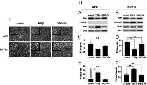
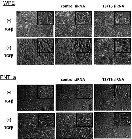
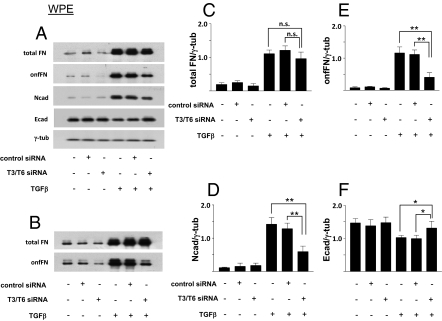
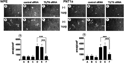
The process termed "epithelial-mesenchymal transition" (EMT) was originally discovered in ontogenic development, and has been shown to be one of the key steps in tumor cell progression and metastasis. Recently, we showed that the expression of some glycosphingolipids (GSLs) is down-regulated during EMT in human and mouse cell lines. Here, we demonstrate the involvement of GalNAc-type (or mucin-type) O-glycosylation in EMT process, induced with transforming growth factor β (TGF-β) in human prostate epithelial cell lines. We found that: (i) TGF-β treatment caused up-regulation of oncofetal fibronectin (onfFN), which is defined by mAb FDC6, and expressed in cancer or fetal cells/tissues, but not in normal adult cells/tissues. The reactivity of mAb FDC6 requires the addition of an O-glycan at a specific threonine, inside the type III homology connective segment (IIICS) domain of FN. (ii) This change is associated with typical EMT characteristics; i.e., change from epithelial to fibroblastic morphology, enhanced cell motility, decreased expression of a typical epithelial cell marker, E-cadherin, and enhanced expression of mesenchymal markers. (iii) TGF-β treatment up-regulated mRNA level of FN containing the IIICS domain and GalNAc-T activity for the IIICS domain peptide substrate containing the FDC6 onfFN epitope. (iv) Knockdown of GalNAc-T6 and T3 inhibited TGF-β-induced up-regulation of onfFN and EMT process. (v) Involvement of GSLs was not detectable with the EMT process in these cell lines. These findings indicate the important functional role of expression of onfFN, defined by site-specific O-glycosylation at IIICS domain, in the EMT process.
Conflict of interest statement
The authors declare no conflict of interest.
Figures


Similar articles
-
Increase of O-glycosylated oncofetal fibronectin in high glucose-induced epithelial-mesenchymal transition of cultured human epithelial cells.PLoS One. 2013 Apr 12;8(4):e60471. doi: 10.1371/journal.pone.0060471. Print 2013. PLoS One. 2013. PMID: 23593224 Free PMC article.
-
Induction of epithelial-mesenchymal transition with O-glycosylated oncofetal fibronectin.FEBS Lett. 2012 Jun 21;586(13):1813-20. doi: 10.1016/j.febslet.2012.05.020. Epub 2012 May 26. FEBS Lett. 2012. PMID: 22641031 Free PMC article.
-
Specific glycosphingolipids mediate epithelial-to-mesenchymal transition of human and mouse epithelial cell lines.Proc Natl Acad Sci U S A. 2009 May 5;106(18):7461-6. doi: 10.1073/pnas.0902368106. Epub 2009 Apr 20. Proc Natl Acad Sci U S A. 2009. PMID: 19380734 Free PMC article.
-
Sweet and sour: the impact of differential glycosylation in cancer cells undergoing epithelial-mesenchymal transition.Front Oncol. 2014 Mar 25;4:59. doi: 10.3389/fonc.2014.00059. eCollection 2014. Front Oncol. 2014. PMID: 24724053 Free PMC article. Review.
-
Role of glycosylation in TGF-β signaling and epithelial-to-mesenchymal transition in cancer.Protein Cell. 2021 Feb;12(2):89-106. doi: 10.1007/s13238-020-00741-7. Epub 2020 Jun 25. Protein Cell. 2021. PMID: 32583064 Free PMC article. Review.
Cited by
-
Concepts of extracellular matrix remodelling in tumour progression and metastasis.Nat Commun. 2020 Oct 9;11(1):5120. doi: 10.1038/s41467-020-18794-x. Nat Commun. 2020. PMID: 33037194 Free PMC article. Review.
-
The Blessed Union of Glycobiology and Immunology: A Marriage That Worked.Medicines (Basel). 2023 Jan 19;10(2):15. doi: 10.3390/medicines10020015. Medicines (Basel). 2023. PMID: 36827215 Free PMC article. Review.
-
Recent Progress on the Role of Fibronectin in Tumor Stromal Immunity and Immunotherapy.Curr Top Med Chem. 2022;22(30):2494-2505. doi: 10.2174/1568026622666220615152647. Curr Top Med Chem. 2022. PMID: 35708087 Review.
-
Site-Specific N- and O-Glycosylation Analysis of Human Plasma Fibronectin.Front Chem. 2021 Jun 15;9:691217. doi: 10.3389/fchem.2021.691217. eCollection 2021. Front Chem. 2021. PMID: 34211961 Free PMC article.
-
Targeting Tn-positive tumors with an afucosylated recombinant anti-Tn IgG.Sci Rep. 2023 Mar 28;13(1):5027. doi: 10.1038/s41598-023-31195-6. Sci Rep. 2023. PMID: 36977722 Free PMC article.
References
-
- Haltiwanger RS, Lowe JB. Role of glycosylation in development. Annu Rev Biochem. 2004;73:491–537. - PubMed
-
- Hakomori S. Traveling for the glycosphingolipid path. Glycoconj J. 2000;17:627–647. - PubMed
-
- Miljan EA, Bremer EG. Regulation of growth factor receptors by gangliosides. Science STKE. 2002;2002:11–10. RE15. - PubMed
-
- Rebbaa A, Chou PM, Bremer EG. Modulation of Growth Factor response in brain tumors by complex carbohydrates. Bull Cancer. 2004;91:E15–E60. - PubMed
-
- Todeschini AR, Dos Santos JN, Handa K, Hakomori SI. Ganglioside GM2-tetraspanin CD82 complex inhibits met and its cross-talk with integrins, providing a basis for control of cell motility through glycosynapse. J Biol Chem. 2007;282:8123–8133. - PubMed
Publication types
MeSH terms
Substances
Grants and funding
LinkOut - more resources
Full Text Sources
Other Literature Sources
Molecular Biology Databases
Miscellaneous
