. 2011 Nov 1;108(44):18049-54.
doi: 10.1073/pnas.1110076108. Epub 2011 Oct 17.

Langerhans cells are precommitted to immune tolerance induction

Affiliations

Langerhans cells are precommitted to immune tolerance induction

Elena Shklovskaya et al. Proc Natl Acad Sci U S A. .

Abstract

Antigen-dependent interactions between T lymphocytes and dendritic cells (DCs) can produce two distinct outcomes: tolerance and immunity. It is generally considered that all DC subsets are capable of supporting both tolerogenic and immunogenic responses, depending on their exposure to activating signals. Here, we tested whether epidermal Langerhans cells (LCs) can support immunogenic responses in vivo in the absence of antigen presentation by other DC subsets. CD4 T cells responding to antigen presentation by activated LCs initially proliferated but then failed to differentiate into effector/memory cells or to survive long term. The tolerogenic function of LCs was maintained after exposure to potent adjuvants and occurred despite up-regulation of the costimulatory molecules CD80, CD86, and IL-12, but was consistent with their failure to translocate the NF-κB family member RelB from the cytoplasm to the nucleus. Commitment of LCs to tolerogenic function may explain why commensal microorganisms expressing Toll-like receptor (TLR) ligands but confined to the skin epithelium are tolerated, whereas invading pathogens that breach the epithelial basement membrane and activate dermal DCs stimulate a strong immune response.


Conflict of interest statement

The authors declare no conflict of interest.

Figures

Fig. 1.
Fig. 1.
Characterization of chimeric mouse models with expression of IE restricted to either LCs or nonepidermal DCs. (A) Schematic representation of LC chimeras. (B) Expression of IE in the skin and skin-draining LNs of chimeric mice. Representative flow cytometric plots are gated to show the frequency of IE+ DCs as a percentage of total DCs. (C) Schematic representation of control chimeras.
Fig. 2.
Fig. 2.
In vivo response of naïve CD4+ T cells to antigen presented by m-LCs or nonepidermal DCs. Fully reconstituted (>3 mo) LC or control chimeras were adoptively transferred with 2 × 10⁵ CFSE-labeled 5C.C7 T cells and s.c. immunized with 10 μg MCC peptide/CFA. (A) Response of donor 5C.C7 T cells in dLNs. Representative flow cytometric plots of CD4 T cells are gated to show the frequency of donor 5C.C7 cells (Left) and their CFSE-dilution profiles (Right). (B) Absolute number of donor 5C.C7 T cells in dLN and spleen. Data are from one representative experiment out of three (3–5 animals per group). (C) Acquisition of effector memory phenotype by donor 5C.C7 T cells. Donor 5C.C7 T cells in dLN were gated for undivided CFSEhigh 5C.C7 cells (gate I) and fully divided CD62LCFSE 5C.C7 cells (gate II). (Right) Mean absolute numbers of cells within the two gates. Values are for one representative experiment. (D and E) Representative flow cytometric plots (D) and absolute number (E) of cytokine-producing donor 5C.C7 cells in dLN. Numbers in D indicate the frequency of cells in each of the four quadrants. (F and G) Antigen presentation by LCs does not support differentiation of CD4+ memory cells. Memory response to day 60 s.c. challenge with peptide/IFA (F) or to day 80 intradermal challenge with peptide-pulsed IE+ splenic DCs (G). Draining LNs were collected 16 h (F) or 3 d after challenge (G). (F Left) Frequency of 5C.C7 cells expressed as a percentage of total CD4 T cells in unchallenged versus challenged mice. (F Center) Expression of CD44 and CD62L. (F Right) Cytokine production after challenge. (G) Frequency of donor 5C.C7 cells (expressed as percent of total CD4 T cells) in draining LNs and ear skin after intradermal ear challenge. One representative experiment out of three is shown.
Fig. 3.
Fig. 3.
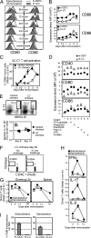
Epicutaneous immunization activates LCs but does not support development of CD4 memory cells. (AC) LC or control chimeras were immunized with HELMCC in cream applied onto hairless abdominal skin. Representative flow profiles (A) and kinetics of CD80 and CD86 expression (B) by m-LCs and m-DDCs in dLN (mean of three per group ± SEM). (C) Activation of 5C.C7 T cells (three mice per group). MFI, mean fluorescence intensity. (D) Expression of CD40, CD80, and CD86 on day 4 after immunization with cream containing either CFA particles, imiquimod, Pam3CSK, poly I:C, lipopolysaccharide (LPS), or curdlan, as indicated. (E) Intracellular IL-12p40/p70 expression by IE+ m-DDCs (control chimeras) and IE+ m-LCs (LC chimeras) in dLN 6 d after epicutaneous immunization with cream containing HELMCC and CFA particles. (Upper) Representative dot plots showing the frequency of IL-12-positive cells (gated) among IE+ m-DCs. (Lower) Absolute number of IL-12+ m-DDCs (circles) and IL-12+ m-LCs (triangles) in dLNs. (F) Frequency of donor 5C.C7 T cells in dLN after memory recall with peptide/IFA on day 90 after epicutaneous immunization of LC chimeras with HELMCC/cream/CFA particulates. (G and H) Response of adoptively transferred 5C.C7 cells in B10.BR mice immunized either epicutaneously for 5 d with HELMCC/cream containing a mixture of CFA particulates, Pam3CSK, poly I:C, imiquimod, and curdlan (triangles) or s.c. with HELMCC/CFA (circles). Absolute numbers of donor 5C.C7 cells (G) and cytokine-producing donor 5C.C7 cells (H) are shown. (I) LCs migrating to draining LNs after immunization fail to translocate the NF-κB subunit RelB to the nucleus. Migratory LCs or DDCs were flow-sorted from dLN of chimeric mice after s.c. (Left) or epicutaneous (Right) immunization, and RelB translocation to the nucleus was analyzed by confocal microscopy. Graphs show the mean percentage (±SEM) of RelB translocations per visual field for 6–8 fields containing >200 DCs per sample.
Fig. 4.
Fig. 4.
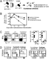
LCs inhibit CD4 T-cell effector responses initiated and maintained by nonepidermal DC subsets. (A) Schematic representation of combined chimeras. (BD) Combined, control, and LC chimeras were adoptively transferred with 2 × 10⁵ CFSE-labeled 5C.C7 T cells and s.c. immunized with pMCC/CFA. (B) Absolute number of donor 5C.C7 T cells in dLN. Data are from one representative experiment of two, with three or four animals per group. (C) Representative flow cytometric plots (Left) and absolute number (Right) of cytokine-producing donor 5C.C7 cells 10 d postimmunization. Each symbol represents an individual mouse. (D) Memory response of combined chimeras to intradermal challenge with peptide-loaded IE+ DCs 80 d after priming. (Left) Frequency of 5C.C7 T cells in draining LN and skin of challenged versus unchallenged mice. (Right) Expression of CD44 and CD62L.

Comment in

  • Inflammation: Under the skin.
    Papatriantafyllou M. Papatriantafyllou M. Nat Rev Immunol. 2011 Nov 4;11(12):800. doi: 10.1038/nri3113. Nat Rev Immunol. 2011. PMID: 22051889 No abstract available.

References

    1. Steinman RM, Banchereau J. Taking dendritic cells into medicine. Nature. 2007;449:419–426. - PubMed
    1. Romani N, Clausen BE, Stoitzner P. Langerhans cells and more: Langerin-expressing dendritic cell subsets in the skin. Immunol Rev. 2010;234:120–141. - PMC - PubMed
    1. Allan RS, et al. Epidermal viral immunity induced by CD8α+ dendritic cells but not by Langerhans cells. Science. 2003;301:1925–1928. - PubMed
    1. Bennett CL, et al. Inducible ablation of mouse Langerhans cells diminishes but fails to abrogate contact hypersensitivity. J Cell Biol. 2005;169:569–576. - PMC - PubMed
    1. Bennett CL, Noordegraaf M, Martina CA, Clausen BE. Langerhans cells are required for efficient presentation of topically applied hapten to T cells. J Immunol. 2007;179:6830–6835. - PubMed

Publication types