

MRI-guided vascular access with an active visualization needle
- PMID: 22006552
- PMCID: PMC3201741
- DOI: 10.1002/jmri.22715
MRI-guided vascular access with an active visualization needle
Abstract
Purpose: To develop an approach to vascular access under magnetic resonance imaging (MRI), as a component of comprehensive MRI-guided cardiovascular catheterization and intervention.
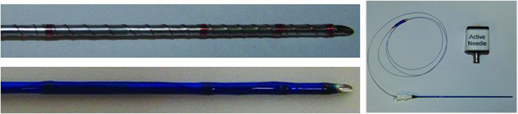
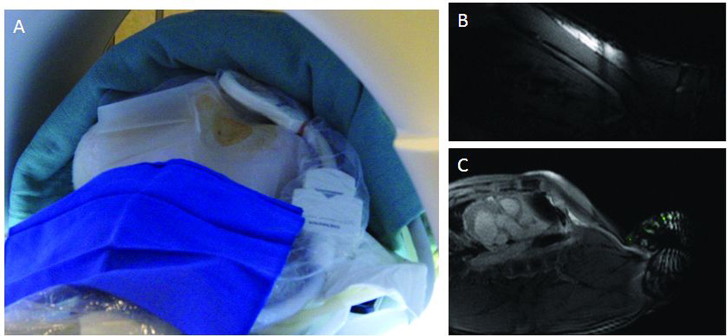
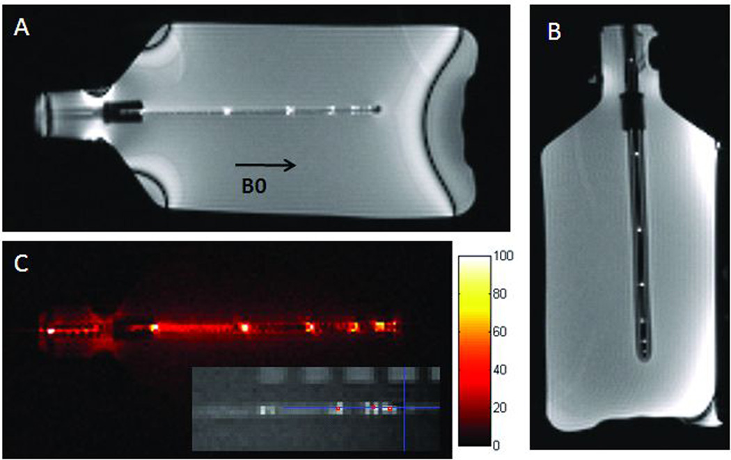
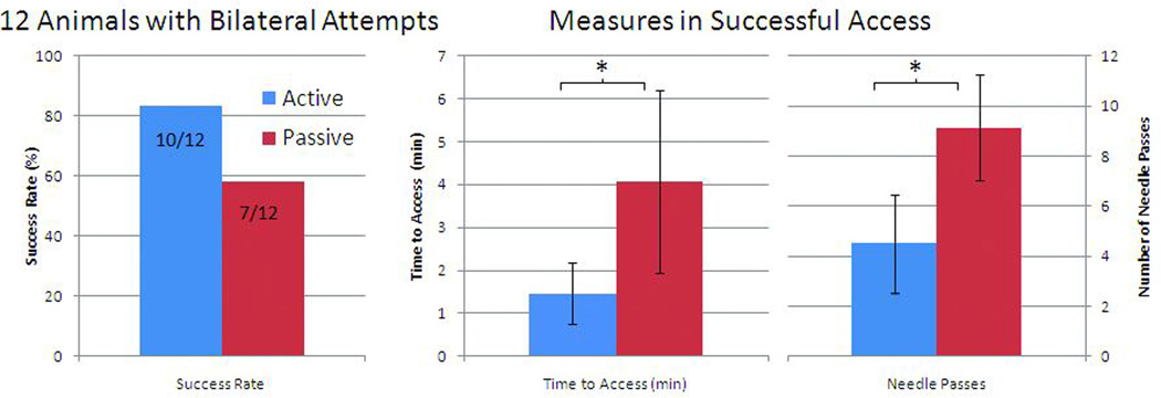
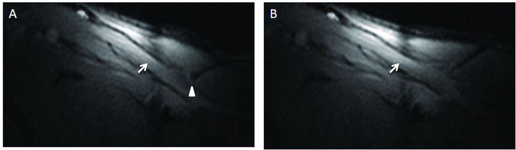
Materials and methods: We attempted jugular vein access in healthy pigs as a model of "difficult" vascular access. Procedures were performed under real-time MRI guidance using reduced field of view imaging. We developed an "active" MRI antenna-needle having an open-lumen, distinct tip appearance and indicators of depth and trajectory in order to enhance MRI visibility during the procedure. We compared performance of the active needle against an unmodified commercial passively visualized needle, measured by procedure success among operators with different levels of experience.
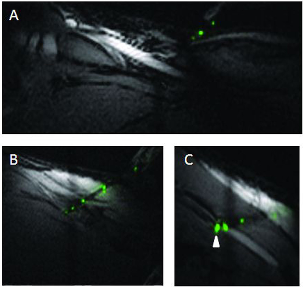
Results: MRI-guided central vein access was feasible using both the active needle and the unmodified passive needle. The active needle required less time (88 vs. 244 sec, P = 0.022) and fewer needle passes (4.5 vs. 9.1, P = 0.028), irrespective of operator experience.
Conclusion: MRI-guided access to central veins is feasible in our animal model. When image guidance is necessary for vascular access, performing this component under MRI will allow wholly MRI-guided catheterization procedures that do not require adjunctive imaging facilities such as x-ray or ultrasound. The active needle design showed enhanced visibility, as expected. These capabilities may permit more complex catheter-based cardiovascular interventional procedures enabled by enhanced image guidance.
Copyright © 2011 Wiley Periodicals, Inc.
Conflict of interest statement
No author has a financial conflict of interest. NHLBI and Siemens have a collaborative research and development agreement covering inteventional cardiovascular MRI.
Figures
References
-
- Samal AK, White CJ. Percutaneous management of access site complications. Catheter Cardiovasc Interv. 2002;57:12–23. - PubMed
-
- Ladd ME, Erhart P, Debatin JF, Romanowski BJ, Boesiger P, McKinnon GC. Biopsy needle susceptibility artifacts. Magn Reson Med. 1996;36:646–651. - PubMed
-
- Frahm C, Gehl HB, Melchert UH, Weiss HD. Visualization of magnetic resonance-compatible needles at 1.5 and 0.2 Tesla. Cardiovasc Intervent Radiol. 1996;19:335–340. - PubMed
-
- Lewin JS, Duerk JL, Jain VR, Petersilge CA, Chao CP, Haaga JR. Needle localization in MR-guided biopsy and aspiration: effects of field strength, sequence design, and magnetic field orientation. AJR Am J Roentgenol. 1996;166:1337–1345. - PubMed
-
- Dumoulin CL, Souza SP, Darrow RD. Real-time position monitoring of invasive devices using magnetic resonance. Magn Reson Med. 1993;29:411–415. - PubMed
Publication types
MeSH terms
Grants and funding
LinkOut - more resources
Full Text Sources
Other Literature Sources
Medical
