Review
. 2011 Nov 28;369(1955):4440-51.
doi: 10.1098/rsta.2011.0261.

Has the time come to use near-infrared spectroscopy as a routine clinical tool in preterm infants undergoing intensive care?

Affiliations
Review

Has the time come to use near-infrared spectroscopy as a routine clinical tool in preterm infants undergoing intensive care?

Gorm Greisen et al. Philos Trans A Math Phys Eng Sci. .

Abstract

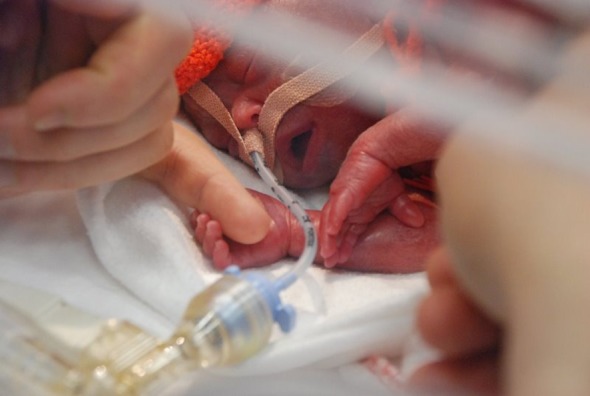
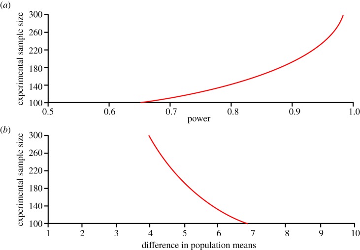
Several instruments implementing spatially resolved near-infrared spectroscopy (NIRS) to monitor tissue oxygenation are now approved for clinical use. The neonatal brain is readily assessible by NIRS and neurodevelopmental impairment is common in children who were in need of intensive care during the neonatal period. It is likely that an important part of the burden of this handicap is due to brain injury induced by hypoxia-ischaemia during intensive care. In particular, this is true for infants born extremely preterm. Thus, monitoring of cerebral oxygenation has considerable potential benefit in this group. The benefit, however, should be weighed against the disturbance to the infant, against the limitations imposed on clinical care and against costs. The ultimate way of demonstrating the 'added value' is by a randomized controlled trial. Cerebral oximetry must reduce the risk of a clinically relevant endpoint, such as death or neurodevelopmental handicap. We estimate that such a trial should recruit about 4000 infants to have the power to detect a reduction in brain injury by one-fifth. This illustrates the formidable task of providing first-grade evidence for the clinical value of diagnostic methods. Is it a window of opportunity for the establishment of a rational basis before another technology is added to an already overly complex newborn intensive care?


Figures

Figure 1.
Figure 1.
An infant shortly after birth born at 25 weeks of gestation. The tube for artificial ventilation passes through the nose to the trachea and is fixed to the skin with tape. The skin is fragile; hence this is acceptable only because ventilation is lifesaving. The cap covering the head and the sheet covering the body are partly to conserve heat, partly for the comfort of the baby, and partly to emphasize the dignity and individuality of the infant. In this context, the practical problem of applying a new probe for monitoring is a significant concern. (Online version in colour.)
Figure 2.
Figure 2.
A clinical management guideline using cerebral oxygenation from the brain to improve the clinical care of extremely preterm infants during the first 48 h after birth. Many aspects of neonatal intensive care are involved. Although the guideline reflects a large body of knowledge of pathophysiology in the newborn brain, some of the interventions have not been shown directly to improve cerebral oxygenation. The guideline was developed during the planning of a randomized controlled trial by a European Academic Consortium with partners from Copenhagen, Utrecht, Leuven, Madrid, Zurich, Uppsala, Milan, Graz, Cambridge, Witten/Herdecke, Lyon and Cork. CPAP, continuous positive airway pressure; GA, gestational age; HB, haemoglobin; PDA, patent ductus arteriosus; PEEP, positive end-expiratory pressure.
Figure 3.
Figure 3.
Calculation of sample size for a randomized controlled trial to demonstrate the clinical benefit of cerebral oximetry in extremely preterm infants. It is hoped that the risk of brain injury can be reduced and it is expected that this will result in improved cognitive development. Calculations are based on an effect size of five points, corresponding to a Cohen d of 0.33, as the standard deviation of a test measuring cognitive function is 15 points, and a study power of 90%. It can be seen that 190 infants will be needed in each of the groups and that the groups must be even larger if the effect size is less, or if the study power should be higher. (Online version in colour.)
Figure 4.
Figure 4.
Magnetic resonance image of the brain shortly after birth in an infant born at 25 weeks of gestation. The brain is immature with few sulci and gyri. The thickness of both the skin and skull is 3–4 mm each. The sylvian fissure is widely open, and there is a large cerebrospinal fluid space in the parieto-occipital region. The fluid spaces constitute significant optical inhomogeneities for near-infrared spectroscopy, because photons can travel far with little chance of absorption or scattering. The result can be large differences in the values of cerebral oxygenation depending on the exact positioning of the source and the detector (MR images courtesy of M. Rutherford, Imperial College, London, UK).
Figure 5.
Figure 5.
The layout of a probe with four sources and two detectors allowing simultaneous measurement of StO2 from four adjacent brain areas. This design may be used for imaging purposes; for the purpose of intensive care, the mean value of StO2 may be used as a global mean. It is less susceptible to imprecision owing to tissue heterogeneity compared with recording from a single brain area. Furthermore, the agreement among the four measures may be used to indicate the reliability of the mean value at any given time. (Online version in colour.)
Figure 6.
Figure 6.
Simulated StO2 in a two-layer geometry as a function of brain SO2. The SO2 in the skin/skull layer was fixed at 70%. All results deviate from the true brain value. The discrepancy is expected for two main reasons. The first is that the spatially resolved spectroscopy (SRS)-based StO2 algorithm ignores the effect of water. The second reason is that the SRS algorithm is based on the assumption of a homogeneous semi-infinite half-space geometry. All designs are responsive to changes in brain SO2. Designs 1 and 2 underread greatly, whereas the designs with larger source–detector distance all produced similar results. (Online version in colour.)
Figure 7.
Figure 7.
The sensitivity of the seven designs against SO2 changes in the skin/scalp layer as indicated by the vertical bars. Brain SO2 was fixed at 70% while the skin/scalp SO2 was varied between 50% and 90%. Simulated StO2 values from the two-layer geometry are influenced by the changes in the skin/scalp layer. In particular, designs 1 and 2 are not robust. (Online version in colour.)

References

    1. Soul J. S., du Plessis A. J.1999New technologies in pediatric neurology. Near-infrared spectroscopy Semin. Pediatr. Neurol. 6101–110.10.1016/S1071-9091(99)80036-9 (doi:10.1016/S1071-9091(99)80036-9) - DOI - DOI - PubMed
    1. Wolf M., Ferrari M., Quaresima V.2007Progress of near-infrared spectroscopy and topography for brain and muscle clinical applications J. Biomed. Opt. 12062104.10.1117/1.2804899 (doi:10.1117/1.2804899) - DOI - DOI - PubMed
    1. Wolf M., Greisen G.2009Advances in near-infrared spectroscopy to study the brain of the preterm and term neonate Clin. Perinatol. 36807–834.10.1016/j.clp.2009.07.007 (doi:10.1016/j.clp.2009.07.007) - DOI - DOI - PubMed
    1. Goldman S., Sutter F., Ferdinand F., Trace C.2004Optimizing intraoperative cerebral oxygen delivery using noninvasive cerebral oximetry decreases the incidence of stroke for cardiac surgical patients Heart Surg. Forum 7E376–E381.10.1532/HSF98.20041062 (doi:10.1532/HSF98.20041062) - DOI - DOI - PubMed
    1. Murkin J., et al. 2007Monitoring brain oxygen saturation during coronary bypass surgery: a randomized, prospective study Anesth. Analg. 10451–58.10.1213/01.ane.0000246814.29362.f4 (doi:10.1213/01.ane.0000246814.29362.f4) - DOI - DOI - PubMed

MeSH terms