. 2011 Dec 9;286(49):42037-42050.
doi: 10.1074/jbc.M111.286948. Epub 2011 Oct 17.

Distinct docking mechanisms mediate interactions between the Msg5 phosphatase and mating or cell integrity mitogen-activated protein kinases (MAPKs) in Saccharomyces cerevisiae

Affiliations

Distinct docking mechanisms mediate interactions between the Msg5 phosphatase and mating or cell integrity mitogen-activated protein kinases (MAPKs) in Saccharomyces cerevisiae

Lorena Palacios et al. J Biol Chem. .

Abstract

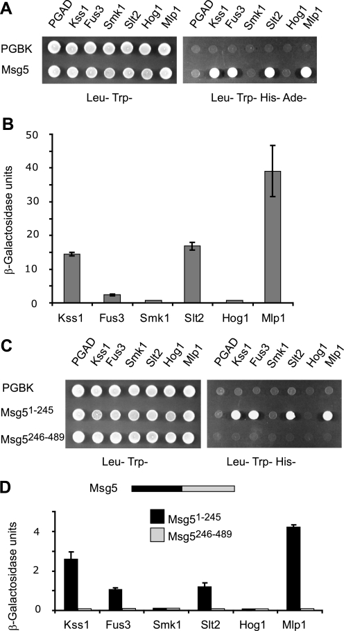
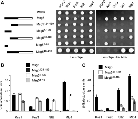
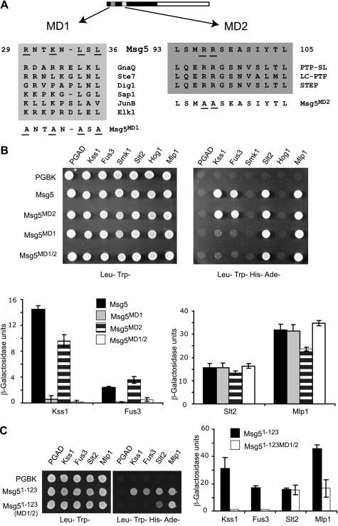
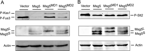
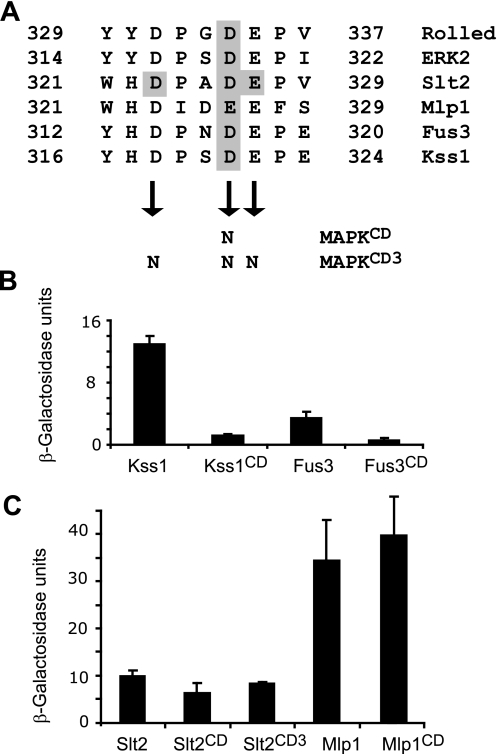
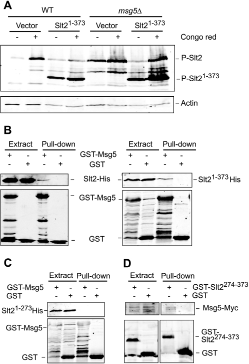
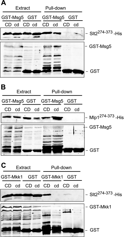
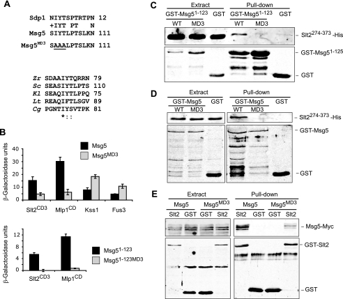
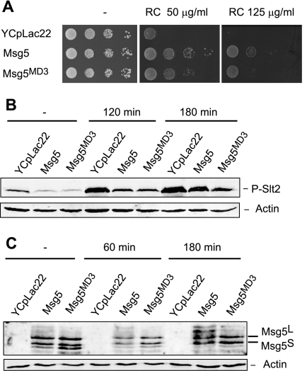
MAPK phosphatases (MKPs) are negative regulators of signaling pathways with distinct MAPK substrate specificities. For example, the yeast dual specificity phosphatase Msg5 dephosphorylates the Fus3 and Slt2 MAPKs operating in the mating and cell wall integrity pathways, respectively. Like other MAPK-interacting proteins, most MKPs bind MAPKs through specific docking domains. These include D-motifs, which contain basic residues that interact with acidic residues in the common docking (CD) domain of MAPKs. Here we show that Msg5 interacts not only with Fus3, Kss1, and Slt2 but also with the pseudokinase Slt2 paralog Mlp1. Using yeast two-hybrid and in vitro interaction assays, we have identified distinct regions within the N-terminal domain of Msg5 that differentially bind either the MAPKs Fus3 and Kss1 or Slt2 and Mlp1. Whereas a canonical D-site within Msg5 mediates interaction with the CD domains of Fus3 and Kss1, a novel motif ((102)IYT(104)) within Msg5 is involved in binding to Slt2 and Mlp1. Furthermore, mutation of this site prevents the phosphorylation of Msg5 by Slt2. This motif is conserved in Sdp1, another MKP that dephosphorylates Slt2, as well as in Msg5 orthologs from other yeast species. A region spanning amino acids 274-373 within Slt2 and Mlp1 mediates binding to this Msg5 motif in a CD domain-independent manner. In contrast, Slt2 uses its CD domain to bind to its upstream activator Mkk1. This binding flexibility may allow MAPK pathways to exploit additional regulatory controls in order to provide fine modulation of both pathway activity and specificity.


Figures

FIGURE 1.
FIGURE 1.
Interaction of Msg5 with MAPKs. A, qualitative analysis of two-hybrid interaction by growth analysis of diploid yeast cells transformed with plasmids encoding the indicated proteins. Vector pGBKT7 (bearing the Gal4 DNA-binding domain) or pGBKT7-Msg5 was transformed into PJ69-4A S. cerevisiae strain and mated with PJ69-4α S. cerevisiae strain transformed with plasmid pGADT7 (bearing the Gal4 activation domain, pGADT7-Kss1, pGADT7-Fus3, pGADT7-Smk1, pGADT7-Slt2, pGADT7-Mlp1, or pGADT7-Hog1. Yeast diploids were spotted onto SD medium lacking leucine and tryptophan (left) or SD medium lacking leucine, tryptophan, histidine, and adenine (right). Cell growth on the latter medium provides evidence for the occurrence of protein-protein interaction. B, semiquantitative analysis of the two-hybrid interaction between Msg5 and the distinct MAPKs based on the level of induction of β-galactosidase. Experiments were performed in triplicate on the diploid strains described in A, with error bars representing S.D. C, qualitative analysis of the two-hybrid interaction between the N-terminal Msg5 (Msg5(1–245)) or C-terminal Msg5 (Msg5(246–489)) region and the different MAPKs. Before mating, pGBKT7, pGBKT7-Msg5(1–245) or pGBKT7-Msg5(246–489) were transformed into PJ69-4A cells, and pGADT7, or pGADT7-Kss1, pGADT7-Fus3, pGADT7-Smk1, pGADT7-Slt2, pGADT7-Mlp1, or pGADT7-Hog1 were transformed into PJ69-4α cells. Two-hybrid assays were performed as indicated in A. D, semiquantitative analysis of the two-hybrid interaction between the same Msg5 protein fragments as in C and the different MAPKs. A schematic showing both halves of Msg5 is also displayed. Experiments were performed in triplicate on the diploid cells described in C, with error bars representing S.D.
FIGURE 2.
FIGURE 2.
Mapping of Msg5 regions that mediate MAPK interaction. A, schematic of full-length and Msg5 fragments analyzed for MAPK interaction (left) and qualitative analysis of the two-hybrid interaction by growth analysis of diploid cells bearing plasmids that encode the indicated proteins or protein fragments (right). Before mating, pGBKT7-Msg5, pGBKT7, pGBKT7-Msg5(126–489), pGBKT7-Msg5(1–123), pGBKT7-Msg5(90–489), pGBKT7-Msg5(1–45), or pGBKT7-Msg5(46–489) were transformed into PJ69-4A cells, and pGADT7 and either pGADT7-Kss1, pGADT7-Fus3, pGADT7-Slt2, or pGADT7-Mlp1 were transformed into PJ69-4α cells. Two-hybrid assays were performed as indicated in Fig. 1A. B and C, semiquantitative analysis of the two-hybrid interaction between Msg5 or the different Msg5 fragments and the distinct MAPKs based on the level of induction of β-galactosidase. Experiments were performed in triplicate on the diploid cells described in A, with error bars representing S.D.
FIGURE 3.
FIGURE 3.
Involvement of distinct docking domains of Msg5 in MAPK binding. A, scheme of Msg5 showing the location of two putative docking domains (MD1 and MD2), which contain conserved amino acid residues (underlined) found in MAPK docking sites of different MAPK-interacting proteins, including yeast Ste7 and Dig1 and mammalian GnaQ, Sap1, JunB, Elk1, PTP-SL, LC-PTP, and STEP. The amino acid substitutions introduced into Msg5 to generate the mutated versions Msg5MD1, Msg5MD2, and Msg5MD1/2 are underlined. B, qualitative (top) and semiquantitative (bottom) analysis of the two-hybrid interaction of diploid cells containing plasmids that encode the indicated proteins. Before mating, pGBKT7, pGBKT7-MSG5, pGBKT7-Msg5MD1, pGBKT7-Msg5MD2, or pGBKT7-Msg5MD1/2 were transformed into PJ69-4A cells, and pGADT7 or pGADT7-Kss1, pGADT7-Fus3, pGADT7-Smk1, pGADT7-Slt2, pGADT7-Mlp1, and pGADT7-Hog1 were transformed into PJ69-4α cells. Two-hybrid assays were performed as indicated in Fig. 1, A and B. C, semi qualitative (left) or semiquantitative (right) analysis of the two-hybrid interaction of diploid cells bearing plasmids that encode the indicated proteins. Before mating, pGBKT7, pGBKT7-Msg5(1–123) or pGBKT7-Msg5(1–123)MD1/2, expressing the first 123 amino acids of Msg5 without or with both MD1 and MD2 substitutions, respectively, were transformed into PJ69-4A cells, and pGADT7, pGADT7-Kss1, pGADT7-Fus3, pGADT7-Slt2, or pGADT7-Mlp1 were transformed into PJ69-4α cells. Two-hybrid assays were performed as indicated in the legend to Fig. 1, A and B.
FIGURE 4.
FIGURE 4.
Effect of mutation of the putative Msg5 docking domains on MAPK phosphorylation. A, Western blot analysis of cell extracts from the DD1-2D (msg5Δ) strain transformed with the empty vector YCplac22, YCplac22MSG5m, YCplac22MSG5MD1m, or YCplac22MSG5MD2m expressing Msg5–6Myc, Msg5MD1-6Myc, or Msg5MD2-6Myc, respectively. Cells were grown to mid-log phase in YPD medium at 24 °C and then treated with 50 nm α-factor for 10 min. B, Western blot analysis of cell extracts from the same strains as in A, after treatment with 30 μg/ml Congo red for 2 h. Analyses were performed using anti-phospho-p44/p42 MAPK (Thr-202/Tyr-204) antibody for immunodetection of phosphorylated Fus3, Kss1, and Slt2 (top), anti-Myc for detecting the two Msg5 isoforms Msg5S and Msg5L (23) (middle), and anti-actin antibodies for actin detection as loading control (bottom). Reproducible results were obtained in different experiments, and selected images correspond to representative blots.
FIGURE 5.
FIGURE 5.
Involvement of the common docking domain of MAPKs on their interaction with Msg5. A, amino acid sequence of the CD domains found in yeast Slt2, Mlp1, Fus3, and Kss1, Drosophila melanogaster Rolled, and mammalian Erk2. Shaded characters indicate the amino acids substituted by asparagines in the different MAPKs (CD and CD3 mutations). B and C, quantitative analysis as indicated in Fig. 1B of the two-hybrid interaction of diploid cells bearing plasmids that encode the indicated proteins. Before mating, pGBKT7, pGBKT7-Msg5 were transformed into PJ69-4A cells, and pGADT7, pGADT7-Kss1, pGADT7-Kss1CD, pGADT7-Fus3, pGADT7-Fus3CD (B), pGADT7-Slt2, pGADT7-Slt2CD, pGADT7-Slt2CD3, pGADT7-Mlp1, or pGADT7-Mlp1CD (C) were transformed into PJ69-4α cells. Error bars, S.D.
FIGURE 6.
FIGURE 6.
Mapping of the Slt2 region that mediates binding to Msg5. A, Western blot analysis of cell extracts from the BY4741 strain (WT) and the isogenic mutant strain Y07373 (msg5Δ) transformed with the vector YCpLac111 or plasmid YCplac111-Slt2(1–373). Cells were grown to mid-log phase in YPD medium at 24 °C (−) and then treated with 30 μg/ml Congo red (CR) for 2 h as indicated. Immunodetection was performed with anti-phospho-p42/44 MAPK (top) and anti-actin (bottom) antibodies as loading control. B and C, Western blot analysis of the in vitro co-purification of recombinant Msg5 with Slt2 and different truncated Slt2 versions. E. coli extracts containing GST or GST-Msg5 were incubated with E. coli extracts containing Slt2-His, Slt2(1–373)-His (B), or Slt2(1–274)-His (C) and glutathione-Sepharose to pull down GST-complexes. Immunodetection was performed using anti-poly-His (top) and anti-GST (bottom) antibodies. D, Western blot analysis of co-purification of Msg5 with Slt2(274–373) fragment. Yeast extracts of the Msg5–6myc-tagged strain YMF1 transformed with the indicated plasmid, pEG(KG) (GST) or pEG(KG)-SLT2(274–373) (GST-SLT2274–373), were incubated with glutathione-Sepharose to pull down GST complexes. Immunodetection was performed with anti-Myc (top) and anti-GST antibodies (bottom). Reproducible results were obtained in different experiments, and selected images correspond to representative blots.
FIGURE 7.
FIGURE 7.
Involvement of the common docking domain of Slt2 and Mlp1 on binding to distinct interactors. A, Western blot analysis of the in vitro co-purification of recombinant Msg5 with the indicated Slt2 fragments. E. coli extracts containing GST or GST-Msg5 were incubated with E. coli extracts containing Slt2(274–373)-His (CD) or Slt2(274–373)(323N,326N,327N)-His (cd) and glutathione-Sepharose to pull down GST complexes. Immunodetection was performed using anti-poly-His (top) and anti-GST (bottom) antibodies. B, Western blot analysis of the in vitro co-purification of recombinant Msg5 with the indicated Mlp1 fragments. E. coli extracts containing GST or GST-Msg5 were incubated with E. coli extracts containing Mlp1(274–373)-His (CD) or Mlp1(274–373)(326N)-His (cd) and glutathione-Sepharose to pull down GST complexes. Immunodetection was performed as in A. C, Western blot analysis of the in vitro co-purification of recombinant Mkk1 with the indicated Slt2 fragments. E. coli extracts containing GST or GST-Mkk1 were incubated with E. coli extracts containing Slt2(274–373)-His (CD) or Slt2(274–373)(323N,326N,327N)-His (cd) and glutathione-Sepharose to pull down GST complexes. Immunodetection was performed as in A. In all cases, reproducible results were obtained in different experiments, and selected images correspond to representative blots.
FIGURE 8.
FIGURE 8.
The MD3 motif mediates binding of Msg5 to Slt2 and Mlp1. A, top, sequence comparison by BLAST analysis between Msg5 and Sdp1 revealed a similar region in the N-terminal part of these two proteins. The area corresponding to the putative Msg5 motif MD3 is shown here. Amino acidic changes introduced in the mutated version Msg5MD3 are underlined. Bottom, multiple alignment of the Msg5 amino acid sequence stretch bearing the MD3 motif with several Msg5 orthologs from different fungi. Alignment was performed by ClustalW (available on the World Wide Web). Identical residues are indicated by asterisks. Dots mark similar residues. Zr, Zygosaccharomyces rouxii; Sc, S. cerevisiae; Kl, Kluyveromyces lactis; Lt, Lachancea thermotolerans; Cg, Candida glabrata. B, semiquantitative analysis of the two-hybrid interaction of diploid cells containing plasmids that encode the indicated proteins. Before mating, pGBKT7, pGBKT7-Msg5, pGBKT7-Msg5MD3, pGBKT7-Msg5(1–123), or pGBKT7-Msg5(1–123)MD3 were transformed into PJ69-4A cells, and pGADT7, pGADT7-Kss1, pGADT7-Fus3, pGADT7-Slt2CD3, or pGADT7-Mlp1CD were transformed into PJ69-4α cells. Two-hybrid assays were performed as indicated in Fig. 1B. C, Western blot analysis of the in vitro co-purification of recombinant GST-Msg5(1–123) (WT) and GST-Msg5(1–123)MD3 (MD3) with Slt2(274–373)-His. E. coli extracts containing GST fusions were incubated with E. coli extracts containing Slt2(274–373)-His and glutathione-Sepharose to pull down GST complexes. Immunodetection was performed using anti-poly-His (top) and anti-GST (bottom) antibodies. D, Western blot analysis of the in vitro co-purification of recombinant GST-Msg5 (WT) and GST-Msg5MD3 (MD3) with Slt2(274–373)-His. E. coli extracts containing GST fusions were incubated with E. coli extracts containing Slt2(274–373)-His and glutathione-Sepharose to pull down GST complexes. Immunodetection was performed as in C. E, Western blot analysis of Msg5–6Myc pulled down by GST-Slt2. Transformants of the yeast DD1-2D strain with YCplac22MSG5m and pEG(KG) (GST) or pEG(KG)-SLT2 (Slt2) were grown to mid-log phase in selective medium at 24 °C, cells were collected, cell extracts were prepared, and GST-Slt2 complexes obtained by purification with glutathione-Sepharose were analyzed by immunoblotting with anti-Myc (top) and anti-GST (bottom) antibodies. In all cases, reproducible results were obtained in different experiments, and selected images correspond to representative blots.
FIGURE 9.
FIGURE 9.
Effect of the lack of Msg5 MD3 motif on the CWI pathway. A, sensitivity to Congo red of msg5Δ (DD1-2D) cells transformed with the vector YCplac22m or plasmid YCplac22MSG5m (Msg5) or YCplac22MSG5MD3m (Msg5MD3). Cells were grown in liquid selective medium at 24 °C, and a 10-fold dilution series of this culture was spotted onto YPD agar medium in the absence (−) or presence of the indicated concentration of Congo red and incubated for 2 days at 24 °C. B, Western blotting analysis of the effect of the Msg5 MD3 mutation on Slt2 phosphorylation. The same transformants as in A were grown to mid-log phase in selective medium at 24 °C, and then aliquots were treated or not with Congo red (30 μg/ml) for 120 and 180 min. Protein extracts were prepared, and phospho-Slt2 and actin (as a loading control) were detected by immunoblot analysis with anti-phospho-p42/44 and anti-actin antibodies, respectively. C, Western blotting analysis of the effect of the Msg5 MD3 mutation on Msg5 phosphorylation. The same transformants as in A were grown to mid-log phase in selective medium at 24 °C, and then aliquots were either left untreated or exposed to Congo red (30 μg/ml) for 60 and 180 min. Proteins extracts were prepared, and Msg5–6Myc or Msg5MD3-6Myc and actin (as a loading control) were detected by immunoblotting analysis with anti-Myc and anti-actin antibodies, respectively. In all cases, reproducible results were obtained in different experiments, and selected images correspond to representative blots.

Similar articles

Cited by

References

    1. Turjanski A. G., Vaqué J. P., Gutkind J. S. (2007) Oncogene 26, 3240–3253 - PubMed
    1. Coulombe P., Meloche S. (2007) Biochim. Biophys. Acta 1773, 1376–1387 - PubMed
    1. Tanoue T., Nishida E. (2002) Pharmacol. Ther. 93, 193–202 - PubMed
    1. Grewal S., Molina D. M., Bardwell L. (2006) Cell. Signal. 18, 123–134 - PMC - PubMed
    1. Bardwell L., Shah K. (2006) Methods 40, 213–223 - PMC - PubMed

Publication types

MeSH terms

LinkOut - more resources