Review
. 2011 Oct 17;195(2):171-81.
doi: 10.1083/jcb.201103171.

Evolution: functional evolution of nuclear structure

Affiliations
Review

Evolution: functional evolution of nuclear structure

Katherine L Wilson et al. J Cell Biol. .

Abstract

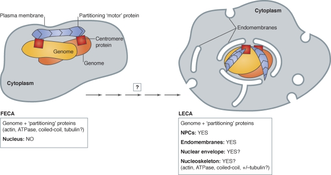
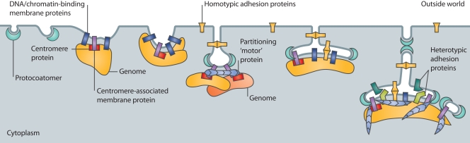
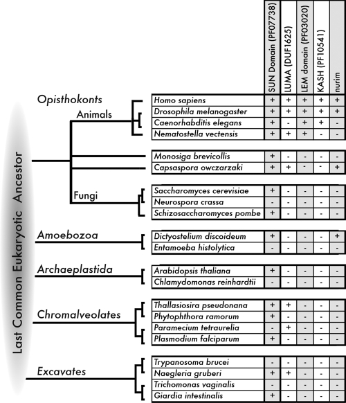
The evolution of the nucleus, the defining feature of eukaryotic cells, was long shrouded in speculation and mystery. There is now strong evidence that nuclear pore complexes (NPCs) and nuclear membranes coevolved with the endomembrane system, and that the last eukaryotic common ancestor (LECA) had fully functional NPCs. Recent studies have identified many components of the nuclear envelope in living Opisthokonts, the eukaryotic supergroup that includes fungi and metazoan animals. These components include diverse chromatin-binding membrane proteins, and membrane proteins with adhesive lumenal domains that may have contributed to the evolution of nuclear membrane architecture. Further discoveries about the nucleoskeleton suggest that the evolution of nuclear structure was tightly coupled to genome partitioning during mitosis.


Figures

Figure 1.
Figure 1.
Proposed incremental transition from FECA (no nuclear structure) to LECA (nucleus). The first eukaryotic common ancestor (FECA) is proposed to have lacked nuclear structure. Partitioning of the duplicated genome (yellow/orange) is proposed to be mediated by the polymerization of protein(s) related to bacterial par “motors” (blue; e.g., actin; ATPase; tubulin; DNA-binding coiled-coil protein), bound to centromere proteins (red squares). Over significant time, the FECA is proposed to have given rise to the last eukaryotic common ancestor (LECA), a cell with fully functional NPCs (not depicted) and endomembranes (Neumann et al., 2010) and, we suggest, a nucleoskeleton that included components involved in genome partitioning. After the LECA, further evolution of nuclear structure followed different pathways as seen in the six living eukaryotic supergroups (Hampl et al., 2009; see Fig. 3).
Figure 2.
Figure 2.
Proposed contributions of membrane proteins to the evolution of nuclear structure. Different types of proteins are proposed to have contributed to the incremental evolution of nuclear structure including soluble proto-coatomer proteins (aqua), DNA- or chromatin-binding membrane proteins (navy blue), centromere- or par system–associated membrane proteins (light purple), homotypic membrane “adhesion” proteins (yellow), and heterotypic membrane adhesion proteins (teal and light green). Blue indicates partitioning proteins (actin, ATPase, DNA-binding coiled-coil protein, tubulin), many of which are components of the nucleoskeleton in living Opisthokonts (see text).
Figure 3.
Figure 3.
Identifying nuclear membrane proteins in diverse eukaryotes. Representatives of five eukaryotic supergroup lineages, queried by BLAST search for potential conservation of open reading frames (ORFs) related to Opisthokont nuclear membrane proteins. Chart indicates whether an ORF(s) homologous to each queried polypeptide was detected (+) or not detected (−) in completed genomes of representative supergroup lineages (Opisthokonts, Amoebozoa, Archaeplastida, Chromalveolates, and Excavates) in GenBank using reciprocal BLAST with e-values > 0.01, as detailed in Table I. ORFs homologous to LUMA and nurim are also present in various bacterial lineages (not depicted). The specific evolutionary relationships between each supergroup and the last eukaryotic common ancestor (LECA) are controversial, as reflected by the lack of specific branching order between these groups. However, the detection of a nuclear-associated homologue (e.g., SUN domain, or LUMA) in more than two supergroups might suggest the homologue was present in the LECA. Negative results are not definitive; for example, we did not recover the S. cerevisiae LEM (“HEH”) domain protein Src1 (see next page). The species M. brevicollis and C. owczarsaki represent the Protist lineage within Opisthokonts and are distantly related to Fungi and Animals.

Similar articles

Cited by

References

    1. Ahmed S., Brickner D.G., Light W.H., Cajigas I., McDonough M., Froyshteter A.B., Volpe T., Brickner J.H.. 2010. DNA zip codes control an ancient mechanism for gene targeting to the nuclear periphery. Nat. Cell Biol. 12:111–118. 10.1038/ncb2011 - DOI - PMC - PubMed
    1. Bengtsson L., Otto H.. 2008. LUMA interacts with emerin and influences its distribution at the inner nuclear membrane. J. Cell Sci. 121:536–548. 10.1242/jcs.019281 - DOI - PubMed
    1. Braunagel S.C., Williamson S.T., Ding Q., Wu X., Summers M.D.. 2007. Early sorting of inner nuclear membrane proteins is conserved. Proc. Natl. Acad. Sci. USA. 104:9307–9312. 10.1073/pnas.0703186104 - DOI - PMC - PubMed
    1. Buch C., Lindberg R., Figueroa R., Gudise S., Onischenko E., Hallberg E.. 2009. An integral protein of the inner nuclear membrane localizes to the mitotic spindle in mammalian cells. J. Cell Sci. 122:2100–2107. 10.1242/jcs.047373 - DOI - PubMed
    1. Cai M., Huang Y., Ghirlando R., Wilson K.L., Craigie R., Clore G.M.. 2001. Solution structure of the constant region of nuclear envelope protein LAP2 reveals two LEM-domain structures: one binds BAF and the other binds DNA. EMBO J. 20:4399–4407. 10.1093/emboj/20.16.4399 - DOI - PMC - PubMed

Publication types