. 2011 Oct 17;195(2):193-201.
doi: 10.1083/jcb.201104076.

High-resolution imaging reveals indirect coordination of opposite motors and a role for LIS1 in high-load axonal transport

Affiliations

High-resolution imaging reveals indirect coordination of opposite motors and a role for LIS1 in high-load axonal transport

Julie Y Yi et al. J Cell Biol. .

Abstract

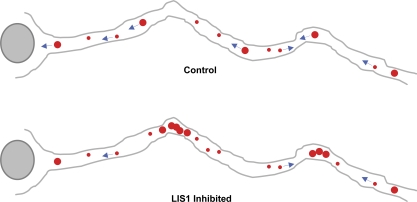
The specific physiological roles of dynein regulatory factors remain poorly understood as a result of their functional complexity and the interdependence of dynein and kinesin motor activities. We used a novel approach to overcome these challenges, combining acute in vivo inhibition with automated high temporal and spatial resolution particle tracking. Acute dynein inhibition in nonneuronal cells caused an immediate dispersal of diverse forms of cargo, resulting from a sharp decrease in microtubule minus-end run length followed by a gradual decrease in plus-end runs. Acute LIS1 inhibition or LIS1 RNA interference had little effect on lysosomes/late endosomes but severely inhibited axonal transport of large, but not small, vesicular structures. Our acute inhibition results argue against direct mechanical activation of opposite-directed motors and offer a novel approach of potential broad utility in the study of motor protein function in vivo. Our data also reveal a specific but cell type-restricted role for LIS1 in large vesicular transport and provide the first quantitative support for a general role for LIS1 in high-load dynein functions.


Figures

Figure 1.
Figure 1.
Rapid dispersal of organelles by acute dynein inhibition. Lysos/LEs rapidly redistribute to the cell periphery by 74.1 Ab microinjection in COS-7 cells within 10 min after injection. (A) Uninjected, IgG, and 74.1 Ab–injected cells are shown at 0 and 10 min after injection. The rightmost panels are time projection images at 1, 5, and 10 min after injection. Kymographs of the indicated regions (dotted rectangles) are shown in the bottom row. Only the 74.1 Ab–injected cell showed en masse lysosome dispersal as shown in the kymograph as plus-biased motion. (B) GFP-NPC1, Rab5-GFP (early endosomes), N-acetylglucosaminyltransferase–GFP (Golgi), and adenovirus Alexa Fluor 546 were dispersed. Dotted circles denote dispersed particles. (C) LysoTracker-positive particles were also dispersed by 70.1 Ab, NudE/L antibody, and the purified CC1 fragment of the p150Glued dynactin protein. (D) N-terminal p150-Glued dynactin antibody, function-blocking LIS1 antibody, and DN LIS1 protein did not affect overall lysosome distribution. All images were taken immediately after injection (0 min) and after 10 min postinjection unless specified otherwise. Bars, 10 µm.
Figure 2.
Figure 2.
High-resolution lyso/LE particle–tracking analysis in COS-7 cells. High-temporal resolution recordings (∼17 frames/s) of lyso/LE motion were processed for analysis. (A) Examples of lysosomal tracks from an uninjected cell showing normal bidirectional lyso/LE motility, a 74.1 Ab–injected cell showing long plus-end–directed travel, and LIS1 DN–injected cells showing normal minus-end–directed transport and oscillatory movement (also see Fig. S2 B). (B–G) Effect of 74.1 and DN LIS1 injection on COS-7 cell lyso/LE motility. Run length (B), percentage of motility (C), and MSD (D) are shown. (E) Net displacement/particle (= [Σ-plus runs (nm) − Σ-minus runs (nm)]/number of particles). (F) Average particle velocity is shown. All error bars represent SEM. *, P < 0.05. (G) Sample size for particle-tracking analysis.
Figure 3.
Figure 3.
Differential lysosomal motility inhibition phenotypes induced by NudE/L and LIS1 inhibition in neurons. Lysos/LEs in rat cortical neurons showed mostly retrograde transport in uninjected and IgG controls, whereas severe inhibition was observed with 74.1 Ab, NudE/L antibody, and LIS1 antibody–injected cells. (A) Kymographs showing cells 1–6 min after injection. Arrest of several large particles is observed by 6 min in LIS1 antibody–injected cells. (B) Time-lapse images of the same LIS1 antibody–injected cell showing large lysosome arresting at the axonal kink (yellow arrow). Boxed areas show LysoTracker red versus dextran fluorescence versus phase-contrast images of the region shown. (C and D) Long-duration videos of the same LIS1 antibody–injected axon and a NudE/L antibody–injected axon. Note the near-complete arrest of motility in the LIS1 antibody–injected case by 20 min as opposed to continued bidirectional motility in the NudE/L antibody–injected case. Note the severe arrest (yellow arrow) as well as the directional reversal (green arrow) in the first 20 min after injection using anti-NudE/L antibody but the extensive bidirectional movement persistent beyond 20 min (orange arrows in C). Lysos/LEs in LIS1 antibody–injected cells showed progressive linear arrest with distance from the cell body (dotted line) during the first 21 min after injection, which is consistent with the diffusion of the antibody along the axon. Although larger particles remained immobile, smaller particles showed rapid transport (orange arrows in D) and could bypass the larger particles (dotted circles). (E) In control cells, lysos/LEs moved mostly in retrograde direction. Kymographs were generated from as near the cell body as imaging could be performed to up to 140 µm away.
Figure 4.
Figure 4.
Knockdown of NudE and LIS1 resembles acute inhibitions. (A) Lysos/LEs in rat hippocampal neurons moved mostly in the retrograde direction in wild-type and scrambled RNAi controls, but an increase in bidirectionally moving lysosomes and severe immobility of larger lysosomal particles (≥1 µm in diameter) were apparent in NudE RNAi and LIS1 RNAi cells, respectively. (A–C) Consistent with the NudE/L antibody–injected case, knockdown of NudE caused an increase in bidirectionally moving lysos/LEs (A and B), whereas knockdown of LIS1 protein more severely interfered with mobility of larger particles than smaller ones, which is consistent with the LIS1 antibody–injected case (A–C). Stationary, anterograde/retrograde, and bidirectional particles were defined as particles that displaced |ΔX| < 1 µm, >5 µm in one direction, and >3 µm in both directions, respectively. The error bars represent SD (*, P < 0.001). The number of particles analyzed are as follows: control (small), n = 60; control (large), n = 55; LIS1 RNAi (small), n = 40; LIS1 RNAi (large), n = 66; NudE RNAi (small), n = 46; and NudE RNAi (large), n = 38 (using 10 axons per condition).
Figure 5.
Figure 5.
Effect of LIS1 inhibition on lyso/LE transport in neurons. Control hippocampal and neocortical neurons exhibited rapid lysos/LEs mostly in the retrograde direction. LIS1 inhibition specifically and potently blocked movement of large lysos/LEs, especially at axonal bends and branch points, with little effect on the smaller vesicular transport. These effects were not observed in nonneuronal cells, which is consistent with a role for LIS1 in transport under higher resistance conditions. Red dots indicate lyso/LE particles. Blue arrows indicate transport directions.

References

    1. Ally S., Larson A.G., Barlan K., Rice S.E., Gelfand V.I. 2009. Opposite-polarity motors activate one another to trigger cargo transport in live cells. J. Cell Biol. 187:1071–1082 10.1083/jcb.200908075 - DOI - PMC - PubMed
    1. Barkus R.V., Klyachko O., Horiuchi D., Dickson B.J., Saxton W.M. 2008. Identification of an axonal kinesin-3 motor for fast anterograde vesicle transport that facilitates retrograde transport of neuropeptides. Mol. Biol. Cell. 19:274–283 10.1091/mbc.E07-03-0261 - DOI - PMC - PubMed
    1. Brady S.T., Pfister K.K., Bloom G.S. 1990. A monoclonal antibody against kinesin inhibits both anterograde and retrograde fast axonal transport in squid axoplasm. Proc. Natl. Acad. Sci. USA. 87:1061–1065 10.1073/pnas.87.3.1061 - DOI - PMC - PubMed
    1. Bremner K.H., Scherer J., Yi J., Vershinin M., Gross S.P., Vallee R.B. 2009. Adenovirus transport via direct interaction of cytoplasmic dynein with the viral capsid hexon subunit. Cell Host Microbe. 6:523–535 10.1016/j.chom.2009.11.006 - DOI - PMC - PubMed
    1. Culver-Hanlon T.L., Lex S.A., Stephens A.D., Quintyne N.J., King S.J. 2006. A microtubule-binding domain in dynactin increases dynein processivity by skating along microtubules. Nat. Cell Biol. 8:264–270 10.1038/ncb1370 - DOI - PubMed

Publication types