. 2011 Dec 1;71(23):7259-69.
doi: 10.1158/0008-5472.CAN-11-1143. Epub 2011 Oct 17.

Lactoferrin-endothelin-1 axis contributes to the development and invasiveness of triple-negative breast cancer phenotypes

Affiliations

Lactoferrin-endothelin-1 axis contributes to the development and invasiveness of triple-negative breast cancer phenotypes

Ngoc-Han Ha et al. Cancer Res. .

Abstract

Triple-negative breast cancer (TNBC) is characterized by the lack of expression of estrogen receptor-α (ER-α), progesterone receptor (PR), and human epidermal growth factor receptor-2 (HER-2). However, pathways responsible for downregulation of therapeutic receptors, as well as subsequent aggressiveness, remain unknown. In this study, we discovered that lactoferrin (Lf) efficiently downregulates levels of ER-α, PR, and HER-2 in a proteasome-dependent manner in breast cancer cells, and it accounts for the loss of responsiveness to ER- or HER-2-targeted therapies. Furthermore, we found that lactoferrin increases migration and invasiveness of both non-TNBC and TNBC cell lines. We discovered that lactoferrin directly stimulates the transcription of endothelin-1 (ET-1), a secreted proinvasive polypeptide that acts through a specific receptor, ET(A)R, leading to secretion of the bioactive ET-1 peptide. Interestingly, a therapeutic ET-1 receptor-antagonist blocked lactoferrin-dependent motility and invasiveness of breast cancer cells. The physiologic significance of this newly discovered Lf-ET-1 axis in the manifestation of TNBC phenotypes is revealed by elevated plasma and tissue lactoferrin and ET-1 levels in patients with TNBC compared with those in ER(+) cases. These findings describe the first physiologically relevant polypeptide as a functional determinant in downregulating all three therapeutic receptors in breast cancer, which uses another secreted ET-1 system to confer invasiveness. Results presented in this article provide proof-of-principle evidence in support of the therapeutic effectiveness of ET-1 receptor antagonist to completely block the lactoferrin-induced motility and invasiveness of the TNBC as well as non-TNBC cells, and thus, open a remarkable opportunity to treat TNBC by targeting the Lf-ET-1 axis using an approved developmental drug.


Figures

Figure 1
Figure 1. Lactoferrin downregulates ERα, PR, and HER-2 in human breast cancer cells
(A) Effect of a single dose of Lf on the levels of indicated proteins in ZR-75 cells. Cells were treated with 20 μg/ml of Lf for indicated times and subjected to Western blot analysis (B) Effect of multiple and increasing doses of Lf on expression levels of proteins in ZR-75 cells. (C) Analysis of ERα, PR and HER-2 levels in ZR-75 cells by confocal microscopy. Cells were treated with 20μg/ml of Lf for 12 hrs, fixed and then stained with protein-specific antibodies. Alexa-488 (ERα and HER-2), Alexa-555 (PR) and DAPI (nucleus). Bar = 5μm.
Figure 2
Figure 2. Lactoferrin downregulates ERα, PR, and HER-2 at a transcriptional level
(A) Northern blot analysis of ERα, PR and HER-2 mRNAs in MCF-7 cells treated with or without 100 μg/ml of Lf for indicated times. (B) MCF-7 cells were treated with or without Lf for indicated time points or with different concentrations of Lf in the presence or absence of proteasomal inhibitor, MG-132. Vinculin was used as a loading control. (C) MDA-MB-231 stable clones expressing ERα or HER-2 were generated as described in the materials and methods. Vinculin was used as a loading control. (D) Effect of Lf on the levels of ERα and HER-2 receptors in MDA-MB-231 pooled clones stably expressing ERα or HER-2.
Figure 3
Figure 3. Lf promotes breast cancer cell migration and invasiveness and compromises anti-invasive activity of tamoxifen and Herceptin
(A) Effect of Lf on cell migration in tamoxifen-treated MCF-7 cells. On the right is the quantification of the average difference in wound closure between 0 and 24 hrs. Left micrographs show representative images of migrated cells after 24 hrs of treatment. (B) Effect of Lf on cell migration in Herceptin-treated MCF-7 cells. On the right is the quantification of the average difference in wound closure between 0 and 24 hrs. Left micrographs show representative images of migrated cells after 24 hrs of treatment. (C) Effect of Lf on cell invasion in tamoxifen-treated MCF-7 cells. E2 was added to the upper wells to enhance tamoxifen effect. (D) Effect of Lf on cell invasion in Herceptin-treated MCF-7 cells. Heregulin was added to the upper wells to enhance Herceptin effect. (E) Effect of Lf on the invasiveness of MDA-MB-231 and MDA-MB-468 cells. Left micrographs are representative images of invaded cells Right is the quantification of the average number of invaded cells. Error bars indicate standard deviation. Scale bars = 0.5mm. *, P < 0.05; **, P < 0.01; ***, P < 0.001
Figure 4
Figure 4. Endothelin-1 is a transcriptional target of Lf
(A & B) Analysis of differentially regulated genes after Lf treatment in MDA-MB-231 and MDA-MB-468 cells by Affymetrix Human Exon 1.0 ST microarrays using Gene-Spring GX 10.0.2. (C) Effect of Lf on ET-1 mRNA levels in breast cancer cells. Actin was used as a loading control. (D) Effect of Lf in the presence or absence of cycloheximide on the induction of ET-1 mRNA in MDA-MB-231 cells.
Figure 5
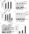
Figure 5. Lf stimulates Endothelin-1 transcription
(A) Effect of Lf on ET-1 promoter activity in breast cancer cells. Cells were treated with 100 μg/ml of Lf for 36 hrs. (B) EMSA analysis of Lf binding to all three Lf-consensus DNA sequences in the ET-1 promoter using the nuclear extracts from MCF-7 cells treated with or without Lf for 6 hrs. (C) Effect of Lf on the ET-1 promoter with wild-type or mutant Lf-binding sequences in MDA-MB-468 cells. (D) EMSA analysis of Lf binding to oligonucleotides encompassing the wild-type Lf-consensus DNA sequence 3 or its three mutant versions using the nuclear extracts from MCF-7 cells treated with or without Lf for 6 hrs. (E) EMSA analysis of Lf-stimulated nuclear extracts from MCF-7 cells in the presence of antibody to Lf. Antibody to rabbit-IgG was used as a negative control (F) Effects of holo- or apo-Lf on the ET-1 promoter luc activity in MDA-MB-468 cells. Error bars indicate standard deviation. *, P < 0.05; **, P < 0.01; ***, P < 0.001
Figure 6
Figure 6. Endothelin-1 receptor antagonist blocks lactoferrin-dependent invasiveness
(A) Concentration of ET-1 in the conditioned media from breast cancer cells treated with or without 100 μg/ml of Lf for 24 hrs as measured by ELISA. (B) Effect of ET-1-receptor inhibitor BQ123 on cell migration of MCF-7 cells treated with or without Lf or ET-1, using Boyden chambers. (C) Effect of ET-1-receptor inhibitor BQ123 on cell invasion of MCF-7 cells treated with or without Lf or ET-1 using Matrigel invasion chambers. *, P < 0.05; **, P < 0.01; ***, P < 0.001
Figure 7
Figure 7. Lf-ET-1 axis promotes TNBC phenotypes
(A) Quantitative PCR analysis of Lf mRNA levels in tumor samples from TNBC and non-TNBC breast cancer patients. (B) Q-PCR analysis of ET-1 mRNAs in TNBC or non-TNBC breast tumors. mRNA experiments were repeated three times, with each point in triplicate. (C) Concentration of Lf and ET-1 in plasma samples from TNBC or non-TNBC patients by ELISA. (D) IHC evaluation of Lf and ET-1 expression in breast tumors from the same patients used in panel B. Table depicts TNBC and non-TNBC tissue samples with scoring of Lf and ET-1 staining intensity: +++ (strong), ++ (medium). (E) Working model for Lf-mediated downregulation of receptors and cell migration/invasion in triple negative breast cancer cells.

Similar articles

Cited by

References

    1. Carey L, Winer E, Viale G, Cameron D, Gianni L. Triple-negative breast cancer: disease entity or title of convenience? Nat Rev Clin Oncol. 2010;12:683–692. - PubMed
    1. Dent R, Trudeau M, Pritchard KI, Hanna WM, Kahn HK, Sawka CA, et al. Triple-negative breast cancer: clinical features and patterns of recurrence. Clin Cancer Res. 2007;15(Pt 1):4429–4434. - PubMed
    1. Rakha EA, El-Sayed ME, Green AR, Lee AH, Robertson JF, Ellis IO. Prognostic markers in triple-negative breast cancer. Cancer. 2007;1:25–32. - PubMed
    1. Sorlie T, Perou CM, Tibshirani R, Aas T, Geisler S, Johnsen H, et al. Gene expression patterns of breast carcinomas distinguish tumor subclasses with clinical implications. Proc Natl Acad Sci U S A. 2001;19:10869–10874. - PMC - PubMed
    1. van’t Veer LJ, Dai H, van de Vijver MJ, He YD, Hart AA, Mao M, et al. Gene expression profiling predicts clinical outcome of breast cancer. Nature. 2002;6871:530–536. - PubMed

Publication types

MeSH terms