. 2011 Nov;138(22):4899-909.
doi: 10.1242/dev.069930. Epub 2011 Oct 17.

The cytoskeletal regulator Genghis khan is required for columnar target specificity in the Drosophila visual system

Affiliations

The cytoskeletal regulator Genghis khan is required for columnar target specificity in the Drosophila visual system

Allison C Gontang et al. Development. 2011 Nov.

Abstract

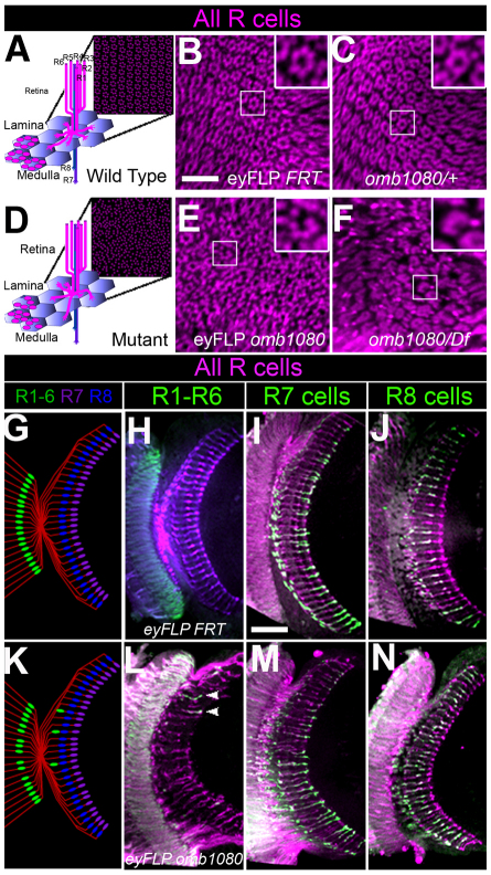
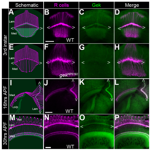
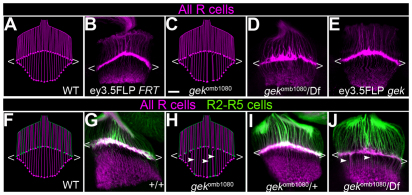
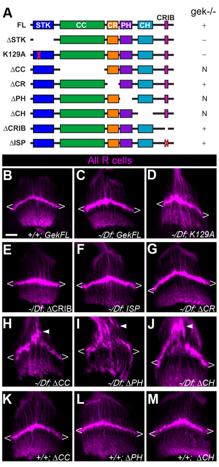
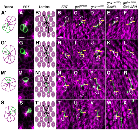
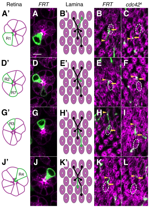
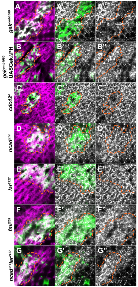
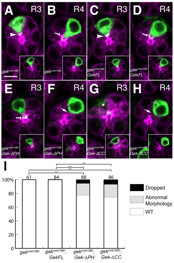
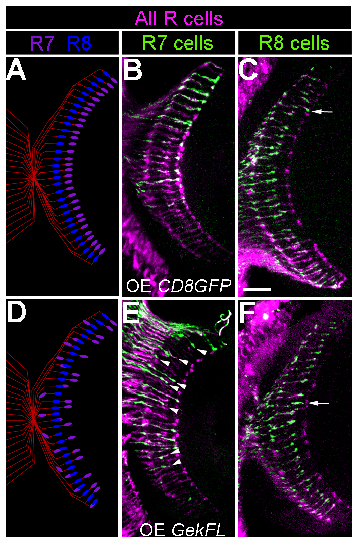
A defining characteristic of neuronal cell type is the growth of axons and dendrites into specific layers and columns of the brain. Although differences in cell surface receptors and adhesion molecules are known to cause differences in synaptic specificity, differences in downstream signaling mechanisms that determine cell type-appropriate targeting patterns are unknown. Using a forward genetic screen in Drosophila, we identify the GTPase effector Genghis khan (Gek) as playing a crucial role in the ability of a subset of photoreceptor (R cell) axons to innervate appropriate target columns. In particular, single-cell mosaic analyses demonstrate that R cell growth cones lacking Gek function grow to the appropriate ganglion, but frequently fail to innervate the correct target column. Further studies reveal that R cell axons lacking the activity of the small GTPase Cdc42 display similar defects, providing evidence that these proteins regulate a common set of processes. Gek is expressed in all R cells, and a detailed structure-function analysis reveals a set of regulatory domains with activities that restrict Gek function to the growth cone. Although Gek does not normally regulate layer-specific targeting, ectopic expression of Gek is sufficient to alter the targeting choices made by another R cell type, the targeting of which is normally Gek independent. Thus, specific regulation of cytoskeletal responses to targeting cues is necessary for cell type-appropriate synaptic specificity.


Figures

Fig. 1.
Fig. 1.
Gek specifically affects the targeting of R1-R6 photoreceptors. (A) Wild-type Drosophila R cell axons from the retina project down into the brain in a common fascicle before extending laterally to innervate specific target columns, forming an array of barrel-like cartridges, each containing six terminals (stained with the R cell-specific mAB24B10, magenta). (B) Adult R cell axon terminals homozygous for a control chromosome. (C) omb1080/+ animals. (D) An omb1080 homozygote. (E) Adult R cell axon terminals homozygous for omb1080. (F) R cell axon terminals in an omb1080/Df animal. Insets in B,C,E,F display single cartridges. (G-J) In adult control eye-specific mosaic animals, R1-R6 cells expressing Rh1-lacZ (green) targeted to the lamina (H), whereas R7 axons labeled with Rh4-lacZ (green) and R8 axons labeled with Rh4-lacZ targeted to single layers in the medulla (I,J). (K-N) Somatic mosaic animals homozygous for omb1080 displayed rare instances in which R1-R6 cells projected into the medulla (L, arrowheads), whereas R7 (M) and R8 (N) targeting was unaffected. Scale bars: 30 μm.
Fig. 2.
Fig. 2.
Gek is enriched in photoreceptor growth cones. (A-D) At the third larval stage in wild type, Gek protein (green) colocalized with R cell axons (mAb24B10, magenta) in both lamina and medulla. (E-H) No specific pattern of Gek expression was detected in a mutant gekNP5192/Df brain. (I-L) Gek expression in wild type 16 hours after puparium formation (APF). (M-P) Gek expression in wild type at 30 hours APF. Ret, retina; Lam, lamina; Med, medulla; Lo/p, lobula and lobula plate. Chevrons delineate the lamina plexus Scale bars: 20 μm.
Fig. 3.
Fig. 3.
gek mutant axons display targeting phenotypes during Drosophila larval development. (A-J) Third instar larval optic lobes, labeling R cells (mAB24B10, magenta) and (F-J) R2-R5 axons (green). (A,C,F,H) Schematics illustrating R cell axon projections into the lamina. Whereas control brains were invariably normal (B,G,I), somatic mosaic animals in which R cells were homozygous for gekomb1080 (E), or transheterozygous for gekomb1080/Df (D,J) displayed axon bundling phenotypes, breaks in the lamina plexus and occasional axons entering the medulla (arrowheads). Chevrons delineate the lamina plexus. Scale bar: 20 μm.
Fig. 4.
Fig. 4.
Structure-function analysis of Gek. (A) Deletion and point mutation constructs. Plus sign denotes constructs that rescue and minus sign denotes constructs that were inactive; N, constructs that caused a novel phenotype. Red cross indicates deletion of the activation (ΔSTK) or binding (ΔCRIB) domain. STK, serine/threonine kinase catalytic domain; CC, coiled-coil domain; CR, cysteine-rich region; PH, pleckstrin homology domain; CH, citron homology domain; CRIB, Cdc42/Rac interactive binding domain. (B-M) Third instar larval optic lobes stained with mAb24B10 (magenta). (B) Full-length gek transgene (GekFL) expression in wild type. (C-J) gekomb1080/Df brains expressing (C) GekFL, (D) Gek-K129A, (E) GekCRIB, (F) GekISP, (G) GekCR, (H) GekCC, (I) GekPH and (J) GekCH. Premature stopping of R cells above the lamina is denoted by arrowheads. (K-M) Expression of GekCC, GekPH or GekCH in wild type. Chevrons delineate the lamina plexus. Scale bar: 20 μm.
Fig. 5.
Fig. 5.
Gek is necessary for columnar targeting of R1-R6 axons. (A′,G′,M′,S′) Schematics of individual ommatidia (magenta) with individual R cell subtypes (green). (A,G,M,S) Confocal images of individual ommatidia (labeled with mAb24B10, magenta) and the control R cell (green). (B′,H′,N′,T′) Schematic projection patterns of one ommatidium. (B,H,N,T) Control R cell axons. (C,I,O,U) gekNP5192 R cell axons. (D,J,P,V) gekomb1080 R cell axons. (E,K,Q,W) gekomb1080 R cell axons that expressed GekFL. (F,L,R,X) gekomb1080 R cell axons that expressed GekPH. Shown are R cell clones (mCD8GFP, green) and target cartridges (mAb24B10, magenta). Arrowheads denote the start of the axon extension and arrow(s) mark the end(s). A dashed line delimits the correct target cartridge. Scale bars: 5 μm.
Fig. 6.
Fig. 6.
Cdc42 is required for columnar targeting. Single-cell MARCM analysis of Cdc42. (A′,D′,G′,J′) Schematics of individual ommatidia (magenta) with individual R cells (green). (A,D,G,J) Individual ommatidia (mAb2410, magenta) and R cells (mCD8GFP, green). (B′,E′,H′,K′) Schematic of the axon projection pattern of one ommatidium. (B,E,H,K) Control R cell axons. (C,F,I,L) Cdc424 R cell axons. Labels are as in Fig. 5.
Fig. 7.
Fig. 7.
Gek and Cdc42 share a common molecular target in R cell growth cones. Phospho-MRLC staining (white) in axon projections of R-cell clones. The clone is labeled in green and axonal projections are labeled with mAb24B10 (magenta). Orange dashed lines indicate the clone border. (A-A″ ) A gekomb1080 clone. (B-B″ ) A gekomb1080 clone expressing GekPH. (C-C″ ) A Cdc424 clone. (D-D″ ) An NcadΔ14 clone. (E-E″ ) A Lar2127 clone. (F-F″ )A flamingoE59 clone. (G-G″ ) An NcadΔ14 Lar2127 clone.
Fig. 8.
Fig. 8.
Gek autoinhibitory function restricts Gek activity to growth cones. (A-H) Ommatidia from gekomb1080 (A,B), gekomb1080; GekFL (C,D), gekomb1080; GekPH (E,F) and gekomb1080; GekCC (G,H) flies. Homozygous R cells are green and the ommatidium magenta (mAb24B10). The bright magenta regions demark developing rhabdomes. (A,C,E,G) R3 cells; (B,D,F,H) R4 cells. Insets display the same cell at a more basal section plane. Normal R3 cells have short apical domains (arrowheads), whereas R4 apical domains protrude centrally (flat-ended arrow). gek mutant R cells expressing these transgenes displayed either abnormally protrusive apical domains (in R3), or lacked apical domains (arrows), or dropped basally within the retina (star). Scale bar: 5 μm. (I) Quantification of mutant phenotypes. Cell bodies were classified as wild-type (WT), abnormal in morphology, or dropped basally. ***, P<0.001 (Fisher’s exact test).
Fig. 9.
Fig. 9.
Overexpression of wild-type Gek alters layer-specific targeting of R7 axons. Transverse sections of the adult medulla stained with mAb24B10 (magenta) and anti-β-galactosidase (green). (A-C) Overexpression of mCD8GFP in all R cells using GlassGal4. (D-F) Overexpression of GekFL using GlassGal4. R7 and R8 cells were labeled with Rh4-lacZ and Rh6-lacZ, respectively. Arrows indicate prematurely terminating axons. Scale bar: 20 μm.

Similar articles

Cited by

References

    1. Berger J., Senti K. A., Senti G., Newsome T. P., Asling B., Dickson B. J., Suzuki T. (2008). Systematic identification of genes that regulate neuronal wiring in the Drosophila visual system. PLoS Genet. 4, e1000085 - PMC - PubMed
    1. Brand A. H., Perrimon N. (1993). Targeted gene expression as a means of altering cell fates and generating dominant phenotypes. Development 118, 401–415 - PubMed
    1. Callahan C. A., Thomas J. B. (1994). Tau-beta-galactosidase, an axon-targeted fusion protein. Proc. Natl. Acad. Sci. USA 91, 5972–5976 - PMC - PubMed
    1. Chen P. L., Clandinin T. R. (2008). The cadherin Flamingo mediates level-dependent interactions that guide photoreceptor target choice in Drosophila. Neuron 58, 26–33 - PMC - PubMed
    1. Chen X. Q., Tan I., Leung T., Lim L. (1999). The myotonic dystrophy kinase-related Cdc42-binding kinase is involved in the regulation of neurite outgrowth in PC12 cells. J. Biol. Chem. 274, 19901–19905 - PubMed

Publication types

MeSH terms

Substances