. 2011 Nov;138(22):4921-30.
doi: 10.1242/dev.070805. Epub 2011 Oct 17.

Protein kinase A acts at the basal body of the primary cilium to prevent Gli2 activation and ventralization of the mouse neural tube

Affiliations

Protein kinase A acts at the basal body of the primary cilium to prevent Gli2 activation and ventralization of the mouse neural tube

Miquel Tuson et al. Development. 2011 Nov.

Abstract

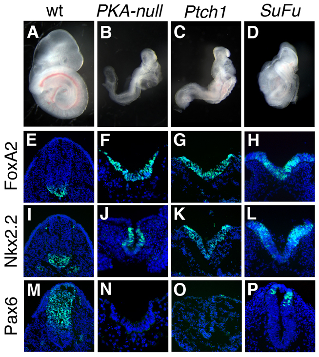
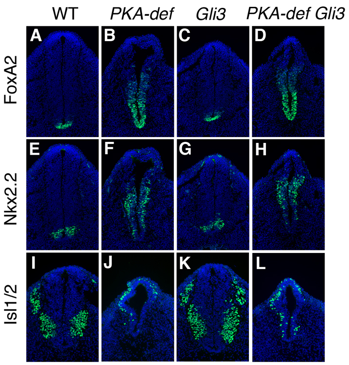
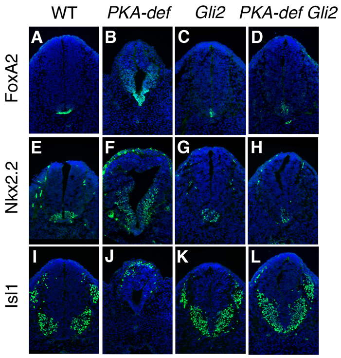
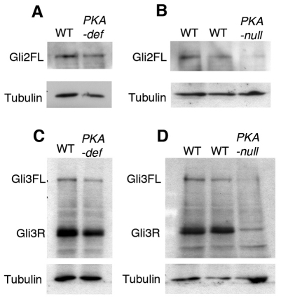
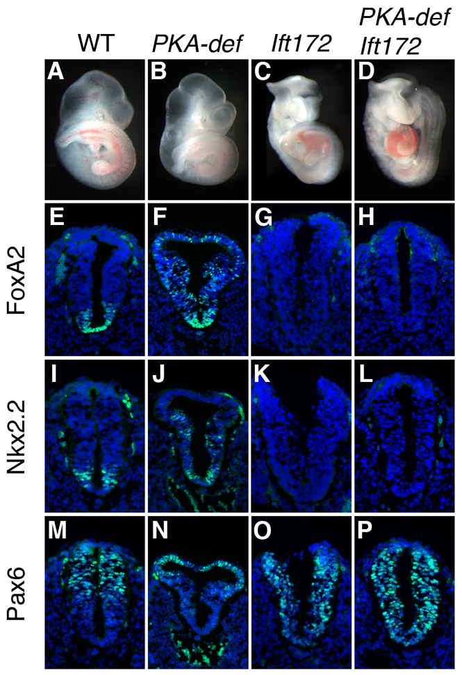
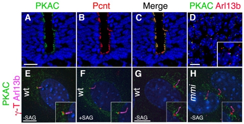
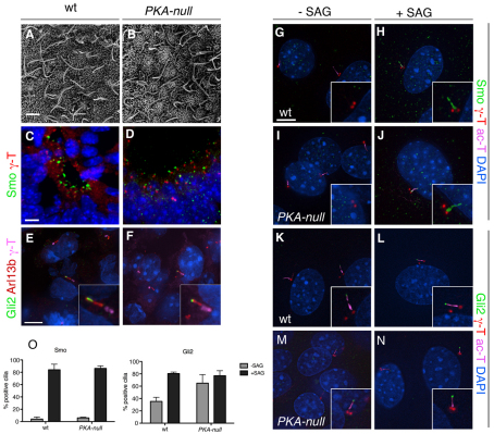
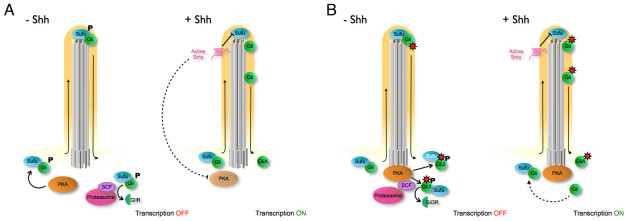
Protein kinase A (PKA) is an evolutionarily conserved negative regulator of the hedgehog (Hh) signal transduction pathway. PKA is known to be required for the proteolytic processing event that generates the repressor forms of the Ci and Gli transcription factors that keep target genes off in the absence of Hh. Here, we show that complete loss of PKA activity in the mouse leads to midgestation lethality and a completely ventralized neural tube, demonstrating that PKA is as strong a negative regulator of the sonic hedgehog (Shh) pathway as patched 1 (Ptch1) or suppressor of fused (Sufu). Genetic analysis shows that although PKA is important for production of the repressor form of Gli3, the principal function of PKA in the Shh pathway in neural development is to restrain activation of Gli2. Activation of the Hh pathway in PKA mutants depends on cilia, and the catalytic and regulatory subunits of PKA are localized to a compartment at the base of the primary cilia, just proximal to the basal body. The data show that PKA does not affect cilia length or trafficking of smoothened (Smo) in the cilium. Instead, we find that there is a significant increase in the level of Gli2 at the tips of cilia of PKA-null cells. The data suggest a model in which PKA acts at the base of the cilium after Gli proteins have transited the primary cilium; in this model the sequential movement of Gli proteins between compartments in the cilium and at its base controls accessibility of Gli proteins to PKA, which determines the fates of Gli proteins and the activity of the Shh pathway.


Figures

Fig. 1.
Fig. 1.
Absence of PKA activity causes maximal activation of the Shh pathway. (A-D) Embryos that lack all four alleles encoding PKA catalytic subunits arrest at E9.0 with a morphology resembling that of Ptch1–/– and Sufu–/– embryos. (E-P) Cross-sections through the open neural plate (thoracic) region of E9.0 wild-type, PKA-null, Ptch1–/– and Sufu–/– embryos, showing a strong ventralization of the neural tube. The expression of FoxA2 (floor plate) (E-H) and Nkx2.2 (V3 progenitors) (I-L) is expanded at all levels of the neural tube, and expression of Pax6 is absent (M-P) (all markers are green). The Sufu–/– embryos express Pax6 in the neural tube, indicating that phenotype is slightly weaker than that of PKA-null and Ptch1–/–. DAPI is blue.
Fig. 2.
Fig. 2.
The neural tube of PKA-deficient Gli3 compound mutants is ventralized. (A-L) Expression of markers of floor plate (FoxA2) (A-D), V3 interneuron progenitors (Nkx2.2) (E-H) and motoneurons (Isl1) (I-L) in cross-sections at the level of the hindlimb of E10.5 wild-type, PKA-deficient, Gli3 single mutant and PKA-deficient Gli3 compound mutant embryos. Markers are in green; DAPI is blue. Whereas Gli3 mutant shows a subtle defects in the caudal neural tube patterning, the floor plate and V3 progenitor domains are expanded, and the motoneuron domain is shifted dorsally in the PKA-deficient Gli3 compound mutants.
Fig. 3.
Fig. 3.
The neural tube phenotype of PKA-deficient Gli2 compound mutant embryos resembles that of Gli2 single mutants. (A-L) Expression of markers of floor plate (FoxA2), V3 interneuron progenitors (Nkx2.2) and motoneurons (Isl1) in cross-sections through at the level of the hindlimb of E10.5 wild-type, PKA-deficient, Gli2 single mutant and PKA-deficient Gli2 compound mutant embryos. Markers are in green; DAPI is blue. In PKA-deficient Gli2 compound mutants, expression of the floor-plate marker FoxA2 is strongly reduced (A-D), Nkx2.2-expressing V3 interneuron progenitors are located in the ventral midline (E,F) and motoneurons span the ventral midline (I-L), as in Gli2 homozygous embryos.
Fig. 4.
Fig. 4.
PKA is required for the stability and processing of Gli proteins. (A,C) Levels of full-length Gli2 and Gli3 are not affected by the partial loss of PKA activity in PKA-deficient embryos, whereas there is a moderate decrease in the level of Gli3R (C). (B,D) Total loss of PKA activity in PKA-null embryos drastically reduces the levels of full-length Gli2 and Gli3, which are almost undetectable, suggesting that PKA is important for the stability of activated full-length Gli2 and Gli3. qPCR experiments showed that RNA levels for Gli3 and Gli2 are reduced 2.2±0.1-fold and 1.7±0.1-fold, respectively, in PKA-null embryos, which is not sufficient to explain the decrease in level of the full-length Gli proteins.
Fig. 5.
Fig. 5.
The primary cilium is required for the effect of PKA mutations on the neural tube. (A-D) PKA-deficient Ift172 compound mutants resemble Ift172 single mutants. (E-P) Expression of neural tube patterning markers FoxA2, Nkx2.2 and Pax6 (in green) in cross-sections through the posterior neural tube, at the position of where the hindlimb is beginning to develop, of E9.5 wild-type, PKA-deficient, Ift172 single and PKA-deficient Ift172 compound mutant embryos. Markers in green; DAPI is blue. PKA-deficient Ift172 compound mutants show the same lack of Hh signaling in the neural tube as Ift172 mutants (compare H with G, L with K, and P with O), indicating that the expansion of ventral neural cell types in PKA-deficient mutants depends on cilia.
Fig. 6.
Fig. 6.
PKA localizes to the basal side of the centrosome in wild-type embryos and MEFs. (A-C) PKA catalytic subunits (PKAC, green) are apically enriched in the E9.5 wild-type neural tube and colocalize with pericentrin (Pcnt) (red). (D) PKA catalytic subunits are localized to the base of cilia (visualized by the cilia marker Arl13b, red) in the mesenchymal cells adjacent to the neural tube of the E10.5 embryo. (E) In MEFs, PKAC (green) is enriched at the base of the primary cilium (Arl13b, pink). PKAC is adjacent to, but does not overlap with, the centrosome marker γ-tubulin (red). (F) PKAC localization to the base of the cilia does not change when Shh pathway is activated in the wild-type MEFs treated with 100 nM SAG for 24 hours. (G,H) PKAC does not accumulate in cilia (Arl13b) of MEFs where retrograde trafficking is blocked by mutation of the retrograde motor Dync2h1mmi. Scale bars: 20 μm in A-C; 10 μm in D; 5 μm in E-H.
Fig. 7.
Fig. 7.
PKA is not required for cilia structure or for localization of Smo, but does affect Gli2 localization to cilia. (A,B) SEM shows that cilia are the same length in the node of E8.0 wild-type (wt) and PKA-null embryos. Wild-type cilia were 3.5±0.5 μm long (n=37); PKA-null cilia were 3.6±0.6 μm long (n=27). (C-F) Localization of Smo and Gli2 in sections of PKA-null embryos. (C,D) Smo (green) localizes to the short cilia of the E9.5 neural plate in wild-type (C) and PKA-null mutant (D) embryos. The base of cilia is marked by γ-tubulin. (E,F) Gli2 is enriched in the cilia of mesenchymal cells surrounding the neural tube in the E9.5 wild-type (E) and PKA-null (F) embryos. Cilia are marked by Arl13b, and the base of cilium is marked by γ-tubulin. (G-N) Localization of Hh pathway protein in wild-type and mutant MEFs. (G-J) Smo localization. Smo is cytoplasmic in unstimulated wild-type MEFs (G) and localizes to the cilia after 24 hours of SAG treatment (H). Smo is not localized to cilia in unstimulated PKA-null MEFs (I) and moves into cilia after 24 hours of SAG treatment (J). (K-N) Gli2 localization. Gli2 is present at wild-type cilia tips in the absence of SAG (K) and is further enriched in cilia tips after SAG treatment. (M,N) Gli2 is enriched in cilia tips in PKA-null MEFs regardless of SAG stimulation. Basal bodies are marked by γ-tubulin and cilia are marked by acetylated α-tubulin. (O) Percentage of Smo+ and Gli2+ cilia in experiments shown in G-N. Smo localization is not affected in PKA-null MEFs, whereas significantly more Gli2 is present in the cilia of PKA-null MEFs than in wild type, in the absence of SAG stimulation (P<0.05). Data are mean±s.e.m. Scale bars: 2 μm in A,B; 5 μm in C-N.
Fig. 8.
Fig. 8.
Two two-compartment models for cilia-dependent regulation of Gli activity. PKA is localized to the base of the cilia, where its function is to prevent activation of the pathway in the absence of ligand, and crucial modifications of Gli protein complexes take place within the cilium. (A) In this model, PKA phosphorylates Gli proteins before they enter the cilium and PKA activity is regulated by Smo. In the absence of ligand, phosphorylation of Gli/Sufu complexes by PKA limits trafficking of the complexes. Phosphorylation of Gli proteins by PKA is also required for processing of Gli repressors by proteasome after they exit the cilium. In the presence of ligand, PKA activity is turned off, perhaps by active Smo in the cilium. Unphosphorylated Gli/Sufu then enters the cilium where the complex dissociates. Full-length free Gli proteins are stabilized and become transcription activators. (B) In this model, PKA phosphorylation is determined by the appropriate Gli substrate, which becomes available only when Gli is complexed with Sufu and only after it has transited the primary cilium. In the absence of Shh, unphosphorylated Gli-Sufu complexes enter the cilium and are modified within the cilium (red sunburst), either via covalent modification or a change in the composition of the complex. After exiting the cilium, Gli2 complexed with Sufu is recognized and phosphorylated by PKA, which stabilizes Gli2/Sufu complex and thereby prevents Gli2 activation and recycling of Gli2 back into the cilium. Phosphorylation by PKA targets Gli3/Sufu complex to the SCF E3 ubiquitin ligase and proteasome for processing into Gli3 repressor. In the presence of Shh ligand, active Smo localizes to the cilia and promotes Gli-Sufu dissociation in the cilium; free Gli proteins are modified in the cilia and are no longer substrates for PKA at the base of cilia. Full-length unphosphorylated Gli proteins can also recycle into the cilium. Smo could, in principle, also have a second activity that turns down PKA activity when the pathway is activated, even if PKA acts on Gli proteins only after they exit the cilium.

Similar articles

Cited by

References

    1. Apionishev S., Katanayeva N. M., Marks S. A., Kalderon D., Tomlinson A. (2005). Drosophila Smoothened phosphorylation sites essential for Hedgehog signal transduction. Nat. Cell Biol. 7, 86–92 - PubMed
    1. Bai C. B., Stephen D., Joyner A. L. (2004). All mouse ventral spinal cord patterning by hedgehog is Gli dependent and involves an activator function of Gli3. Dev. Cell 6, 103–115 - PubMed
    1. Barzi M., Berenguer J., Menendez A., Alvarez-Rodriguez R., Pons S. (2010). Sonic-hedgehog-mediated proliferation requires the localization of PKA to the cilium base. J. Cell Sci. 123, 62–69 - PubMed
    1. Besschetnova T. Y., Kolpakova-Hart E., Guan Y., Zhou J., Olsen B. R., Shah J. V. (2010). Identification of signaling pathways regulating primary cilium length and flow-mediated adaptation. Curr. Biol. 20, 182–187 - PMC - PubMed
    1. Borland G., Smith B. O., Yarwood S. J. (2009). EPAC proteins transduce diverse cellular actions of cAMP. Br. J. Pharmacol. 158, 70–78 - PMC - PubMed

Publication types

MeSH terms