. 2011 Nov;138(22):4931-42.
doi: 10.1242/dev.066597. Epub 2011 Oct 17.

HESX1- and TCF3-mediated repression of Wnt/β-catenin targets is required for normal development of the anterior forebrain

Affiliations

HESX1- and TCF3-mediated repression of Wnt/β-catenin targets is required for normal development of the anterior forebrain

Cynthia L Andoniadou et al. Development. 2011 Nov.

Abstract

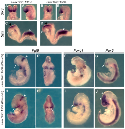
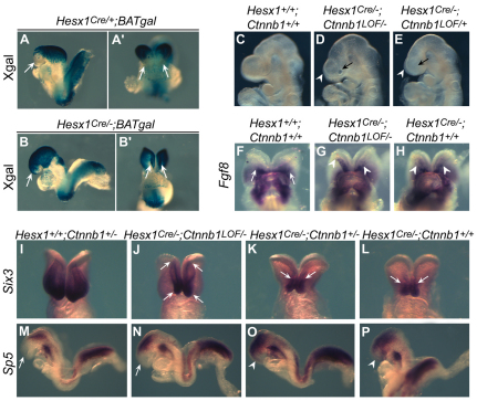
The Wnt/β-catenin pathway plays an essential role during regionalisation of the vertebrate neural plate and its inhibition in the most anterior neural ectoderm is required for normal forebrain development. Hesx1 is a conserved vertebrate-specific transcription factor that is required for forebrain development in Xenopus, mice and humans. Mouse embryos deficient for Hesx1 exhibit a variable degree of forebrain defects, but the molecular mechanisms underlying these defects are not fully understood. Here, we show that injection of a hesx1 morpholino into a 'sensitised' zygotic headless (tcf3) mutant background leads to severe forebrain and eye defects, suggesting an interaction between Hesx1 and the Wnt pathway during zebrafish forebrain development. Consistent with a requirement for Wnt signalling repression, we highlight a synergistic gene dosage-dependent interaction between Hesx1 and Tcf3, a transcriptional repressor of Wnt target genes, to maintain anterior forebrain identity during mouse embryogenesis. In addition, we reveal that Tcf3 is essential within the neural ectoderm to maintain anterior character and that its interaction with Hesx1 ensures the repression of Wnt targets in the developing forebrain. By employing a conditional loss-of-function approach in mouse, we demonstrate that deletion of β-catenin, and concomitant reduction of Wnt signalling in the developing anterior forebrain of Hesx1-deficient embryos, leads to a significant rescue of the forebrain defects. Finally, transcriptional profiling of anterior forebrain precursors from mouse embryos expressing eGFP from the Hesx1 locus provides molecular evidence supporting a novel function of Hesx1 in mediating repression of Wnt/β-catenin target activation in the developing forebrain.


Figures

Fig. 1.
Fig. 1.
hesx1 and hdl (tcf3a) genetically interact for proper anterior-posterior patterning of the zebrafish brain. Lateral views of 2 day post-fertilisation zebrafish embryos. (A,B) Untreated embryos of hdl+/– control (A) and hdl–/– (B) genotypes, both showing normal forebrain development. (C,D) Injection with zebrafish hesx1 MO at the one-cell stage to knockdown Hesx1 results in anterior defects in hdl–/– embryos (arrowhead in D), which are sensitised to increased levels of Wnt signalling. Injection of hesx1 MO into control hdl+/– embryos has no effect (C). (E,F) Anterior defects generated by hesx1 MO injection are rescued by co-injection of murine Hesx1 mRNA (F). Injection of Hesx1 mRNA alone has no effect (E).
Fig. 2.
Fig. 2.
Gene dosage-dependent forebrain defects in mouse embryos genetically deficient for Hesx1 and Tcf3. In situ hybridisation with Fgf8, Foxg1 and Pax6 antisense riboprobes on 9.5 dpc embryos. (A,A′) Fgf8 expression in the wild-type brain is restricted to the anterior neural ridge (ANR, arrows) and at the mid-hindbrain boundary (MHB, arrowhead). Lateral view in A, frontal in A′. (B) Foxg1 is expressed in the normal developing telencephalic vesicles (arrows). (C) In the wild-type brain, Pax6 is expressed in the dorsal telencephalon (arrow), posterior forebrain (arrowheads) and eye. (D,D′) In Hesx1Cre/+;Tcf3+/– double heterozygotes, Fgf8 expression in the MHB (arrowheads) is normal but is reduced in the ANR (arrows). (E) Foxg1 expression in the telencephalon is severely reduced in Hesx1Cre/+;Tcf3+/– embryos (arrows). (F) Pax6 expression in the telencephalon and eye is decreased but expression in the posterior forebrain is normal (arrowheads). (G-I) Forebrain defects are very severe in Hesx1Cre/–;Tcf3+/– embryos and most of the forebrain is missing, as evidenced by the lack of Fgf8 (G,G′) and Foxg1 (H) expression. Note the normal expression of Fgf8 in the MHB (arrowhead in G) and the minimal expression of Pax6 (arrowhead in I).
Fig. 3.
Fig. 3.
Tcf3 is required within the anterior neuroectoderm for normal mouse forebrain development. (A-D) In situ hybridisation with Six3 and Sp5 antisense riboprobes on 8.5 dpc Hesx1Cre/+;Tcf3fl/– (B,B′) and control Hesx1Cre/+;Tcf3+/+ (A,A′) embryos. Expression of Six3, a marker of the anterior forebrain primordium in 8.5 dpc embryos, is reduced in the Hesx1Cre/+;Tcf3fl/– mutant compared with the control. By contrast, the expression domain of Sp5, a direct Wnt/β-catenin target gene, is rostrally expanded into the prospective forebrain region of the Hesx1Cre/+;Tcf3fl/– mutant (arrow in D) as compared with a control embryo, which does not express Sp5 in the prospective forebrain (arrow in C). The arrowheads in C and D denote Sp5 expression in the midbrain (black) and tailbud (white). (E-J) In situ hybridisation with Fgf8, Foxg1 and Pax6 antisense riboprobes on 9.5 dpc Hesx1Cre/+;Tcf3fl/– embryos with severe (class IV) or mild (class I-III) forebrain defects. Stage-matched wild-type controls are shown in Fig. 2A-C. (E,E′) Fgf8 expression at the ANR is severely reduced (arrow in E), with only a minimal domain of expression remaining (arrow in E′); however, expression at the MHB remains normal (arrowhead in E). In an embryo with severe anterior truncation (F), Foxg1 expression, which is normally at the telencephalic vesicles, is lost. Similarly, the anterior Pax6 expression domain is almost absent due to the loss of forebrain tissue and only a small patch of Pax6-positive cells, probably corresponding to posterior forebrain, is detectable (arrow in G). In mildly affected Hesx1Cre/+;Tcf3fl/– mutants, the Fgf8 expression domain at the ANR is reduced and restricted to the midline (arrows in H,H′), but MHB expression is unaffected (arrowhead in H). Foxg1 expression in the telencephalon is severely reduced in a mildly affected Hesx1Cre/+;Tcf3fl/– embryo (arrow in I). In these embryos, Pax6 expression in the telencephalon and eye is decreased but expression in the posterior forebrain is normal (arrowheads in J).
Fig. 4.
Fig. 4.
Loss of function of β-catenin is sufficient to improve forebrain patterning in mouse Hesx1Cre/–;Ctnnb1LOF/– embryos. (A,A′) X-Gal staining reveals BATgal activity in the neural plate of Hesx1Cre/+;BATgal control embryo at 8.5 dpc, but the anteriormost forebrain is not stained (arrows). (B,B′) By contrast, in the Hesx1Cre/– mutant, the anterior forebrain is BATgal positive, suggesting ectopic activation of the Wnt/β-catenin signalling pathway (arrows). (C-E) The conditional inactivation of β-catenin in a Hesx1Cre/–;Ctnnb1LOF/– embryo (D) leads to a significant improvement of telencephalic (arrowhead) and eye (arrow indicating the presence of an optic vesicle) development compared with a Hesx1Cre/–;Ctnnb1LOF/+ embryo (E). However, compared with a wild-type control embryo (C), this is not a full restoration to normal development. (F-H) Frontal views of embryos after in situ hybridisation with antisense riboprobes against Fgf8. Normal expression at the ANR of a wild-type embryo at 3-5 somites (arrows in F). In Hesx1Cre/– mutants, the expression of Fgf8 at the ANR is reduced and restricted to the midline (arrowheads in H). In the Hesx1Cre/–;Ctnnb1LOF/– embryo (G) there is an asymmetric expansion in Fgf8 expression compared with the homozygous Hesx1 mutant (H). Arrowheads indicate the limit of Fgf8 expression, with broader expression on the right-hand side. An asymmetric improvement of forebrain defects in Hesx1Cre/–;Ctnnb1LOF/– embryos is often seen at later stages. (I-P) In situ hybridisation with antisense riboprobes against Six3 and Sp5 on 8.5 dpc embryos at 8-10 somites. The Six3 expression domain in the anterior forebrain of the Hesx1Cre/–;Ctnnb1LOF/– embryo (arrows in J) is larger than in Hesx1Cre/–;Ctnnb1+/– and Hesx1Cre/– mutants (arrows in K,L), but smaller than in the control embryo (I). Expression of the Wnt/β-catenin direct target gene Sp5 is normally excluded from the anterior forebrain (arrow in M), and it remains so in the Hesx1Cre/–;Ctnnb1LOF/– embryo (arrow in N). However, ectopic Sp5 expression is detected in the anterior forebrain of Hesx1Cre/–;Ctnnb1+/– and Hesx1Cre/– mutants (arrowheads in O,P).
Fig. 5.
Fig. 5.
Purification of anterior forebrain precursors by flow sorting from Hesx1eGFP/+ and Hesx1eGFP/eGFP mouse embryos. (A) Targeting strategy for the generation of the Hesx1-eGFP allele. Top to bottom: structure of the murine Hesx1 locus; Hesx1-eGFP targeting vector; targeted allele prior to and after flipase-mediated excision of the Neo cassette; expected bands for the targeted and wild-type alleles after Southern blot analysis of DNA samples digested with EcoRI and hybridised with an external probe (red line in top schematic). The DTA cassette is irrelevant to this study as it has not been activated in the presence of Cre in the experiments presented here. (B) Southern blot analysis of wild-type (Hesx1+/+) and Hesx1eGFP/+ ES cell clones digested with EcoRI and hybridised with an external probe (red line in A). Only the 4.3 kb wild-type band is detected in the Hesx1+/+ sample, whereas both wild-type and mutant (3.9 kb) bands are detected in three correctly targeted Hesx1eGFP/+ clones. (C) Dorsal view of the neural plate of a 3-somite stage Hesx1eGFP/+ embryo showing eGFP fluorescence in the anterior forebrain primordium during normal development (arrowheads). (D) In the Hesx1eGFP/eGFP embryo, in which there is no Hesx1 expression, the anterior forebrain domain marked by eGFP fluorescence becomes medially restricted (arrowheads). (E) Flow sorting of dissociated whole embryos between 3 and 5 somites allows the specific isolation of cells from the prospective anterior forebrain through eGFP fluorescence from the Hesx1 locus. Scatter plots from a representative experiment are shown. Purified cells from heterozygous Hesx1eGFP/+ (normal) or homozygous Hesx1eGFP/eGFP mutant embryos were used for RNA isolation and subsequent microarray analysis.

Similar articles

Cited by

References

    1. Andoniadou C. L., Signore M., Sajedi E., Gaston-Massuet C., Kelberman D., Burns A. J., Itasaki N., Dattani M., Martinez-Barbera J. P. (2007). Lack of the murine homeobox gene Hesx1 leads to a posterior transformation of the anterior forebrain. Development 134, 1499–1508 - PMC - PubMed
    1. Brannon M., Brown J. D., Bates R., Kimelman D., Moon R. T. (1999). XCtBP is a XTcf-3 co-repressor with roles throughout Xenopus development. Development 126, 3159–3170 - PubMed
    1. Brantjes H., Roose J., van de Wetering M., Clevers H. (2001). All Tcf HMG box transcription factors interact with Groucho-related co-repressors. Nucleic Acids Res. 29, 1410–1419 - PMC - PubMed
    1. Brault V., Moore R., Kutsch S., Ishibashi M., Rowitch D. H., McMahon A. P., Sommer L., Boussadia O., Kemler R. (2001). Inactivation of the beta-catenin gene by Wnt1-Cre-mediated deletion results in dramatic brain malformation and failure of craniofacial development. Development 128, 1253–1264 - PubMed
    1. Braun M. M., Etheridge A., Bernard A., Robertson C. P., Roelink H. (2003). Wnt signaling is required at distinct stages of development for the induction of the posterior forebrain. Development 130, 5579–5587 - PubMed

Publication types

MeSH terms