

Epidemiology, diagnosis, and management of cystic lesions of the pancreas
- PMID: 22007199
- PMCID: PMC3191780
- DOI: 10.1155/2012/147465
Epidemiology, diagnosis, and management of cystic lesions of the pancreas
Abstract
Although little is known on the true prevalence of pancreatic cysts, physicians are currently more frequently confronted with pancreatic cysts because of the increasing use of sophisticated cross-sectional abdominal imaging. Cystic lesions of the pancreas comprise of a heterogeneous group of diagnostic entities, some of which are benign such as inflammatory pseudocysts or serous cystadenomas and do not require resection when asymptomatic. Others like mucinous cysts or intraductal papillary mucinous neoplasms (IPMN) have a malignant potential and in these cases surgical resection is often indicated. For this reason an adequate distinction between the various cysts is crucial to optimize management strategy. Different diagnostic methods that could be of value in the differentiation include radiologic imaging techniques such as CT, MR, and endosonography. In addition, fluid aspiration for cytopathology, tumormarkers or molecular analysis is widely used. Different guidelines are available but so far no optimal diagnostic algorithm exists. We summarize the epidemiology, classification, clinical presentation, diagnostics, management, and future perspectives.
Figures
References
-
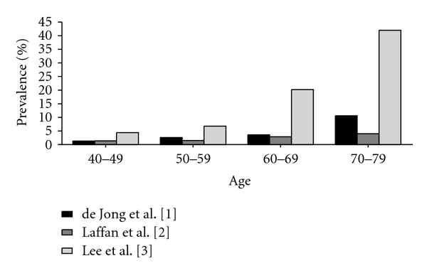
- de Jong K, Nio CY, Hermans JJ, et al. High prevalence of pancreatic cysts detected by screening magnetic resonance imaging examinations. Clinical Gastroenterology and Hepatology. 2010;8(9):806–811. - PubMed
-
- Lee KS, Sekhar A, Rofsky NM, Pedrosa I. Prevalence of incidental pancreatic cysts in the adult population on MR imaging. American Journal of Gastroenterology. 2010;105(9):2079–2084. - PubMed
-
- Zhang XM, Mitchell DG, Dohke M, Holland GA, Parker L. Pancreatic cysts: depiction on single-shot fast spin-echo MR images. Radiology. 2002;223(2):547–553. - PubMed
LinkOut - more resources
Full Text Sources
