

The effect of pulling out cochlear implant electrodes on inner ear microstructures: a temporal bone study
- PMID: 22007225
- PMCID: PMC3191782
- DOI: 10.1155/2011/107176
The effect of pulling out cochlear implant electrodes on inner ear microstructures: a temporal bone study
Abstract
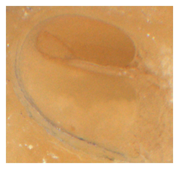
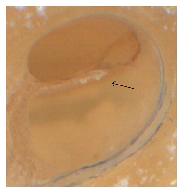
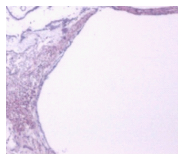
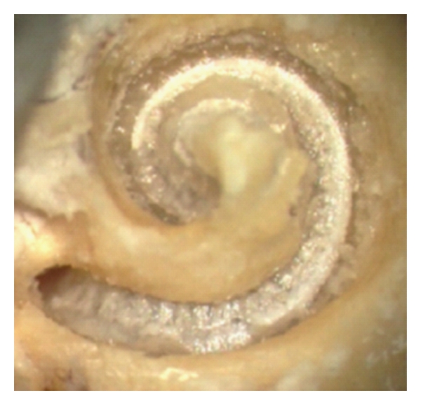
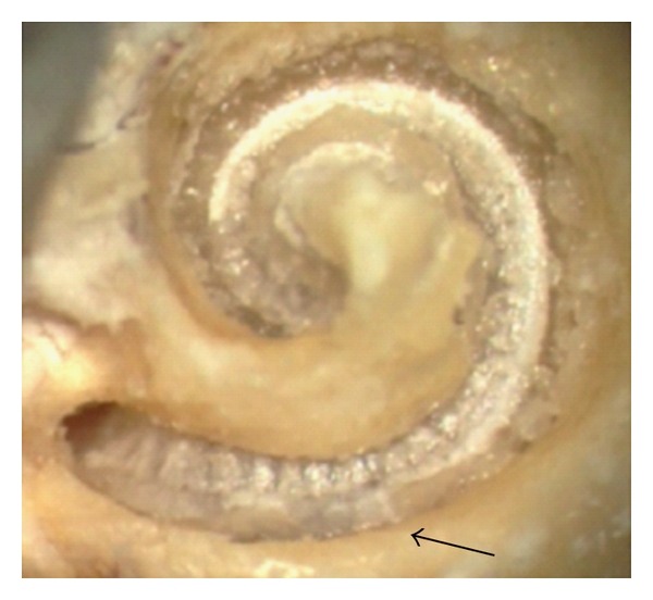
The exchange of an cochlear implant or the re-positioning of an electrode have become more frequently required than a decade ago. The consequences of such procedures at a microstructural level within the cochlea are not known. It was the aim of the present study to further investigate the effects of an CI electrode pull-out. Therefore 10 freshly harvested temporal bones (TB) were histologically evaluated after a cochlear implant electrode pull-out of a perimodiolar electrode. In additional 9 TB the intrascalar movements of the CI electrode while being pulled-out were digitally analysed by video- capturing. Histologically, a disruption of the modiolar wall or the spiral osseous lamina were not observed. In one TB, a basilar membrane lifting up was found, but it could not be undoubtedly attributed to the pull-out of the electrode. When analyzing the temporal sequence of the electrode movement during the pull-out, the electrode turned in one case so that the tip elevates the basilar membrane. The pull- out of perimodiolarly placed CI electrodes does not damage the modiolar wall at a microstructural level and should be guided (e.g., forceps) to prevent a 90 o turning of the electrode tip into the direction of the basilar membrane.
Figures
Similar articles
-
Factors associated with incomplete insertion of electrodes in cochlear implant surgery: a histopathologic study.Audiol Neurootol. 2011;16(2):69-81. doi: 10.1159/000316445. Epub 2010 Jun 19. Audiol Neurootol. 2011. PMID: 20571258 Free PMC article.
-
Perimodiolar electrodes in cochlear implant surgery.Acta Otolaryngol. 2001 Jan;121(2):216-9. doi: 10.1080/000164801300043569. Acta Otolaryngol. 2001. PMID: 11349782
-
Combining perimodiolar electrode placement and atraumatic insertion properties in cochlear implantation -- fact or fantasy?Acta Otolaryngol. 2006 May;126(5):475-82. doi: 10.1080/00016480500437393. Acta Otolaryngol. 2006. PMID: 16698696
-
An overview of cochlear implant electrode array designs.Hear Res. 2017 Dec;356:93-103. doi: 10.1016/j.heares.2017.10.005. Epub 2017 Oct 18. Hear Res. 2017. PMID: 29102129 Review.
-
Optimal electrode design: Straight versus perimodiolar.Eur Ann Otorhinolaryngol Head Neck Dis. 2016 Jun;133 Suppl 1:S63-5. doi: 10.1016/j.anorl.2016.04.014. Epub 2016 May 30. Eur Ann Otorhinolaryngol Head Neck Dis. 2016. PMID: 27256964 Review.
Cited by
-
Why Pre-Curved Modiolar Hugging Electrodes Only Cover The Basal Turn of The Cochlea and Not Beyond that?J Int Adv Otol. 2018 Dec;14(3):376-381. doi: 10.5152/iao.2018.5831. J Int Adv Otol. 2018. PMID: 30644378 Free PMC article.
-
Advances to electrode pullback in cochlear implant surgery.ScientificWorldJournal. 2012;2012:126767. doi: 10.1100/2012/126767. Epub 2012 Oct 22. ScientificWorldJournal. 2012. PMID: 23193376 Free PMC article.
-
The Pull-Back Technique for the 532 Slim Modiolar Electrode.Biomed Res Int. 2019 May 23;2019:6917084. doi: 10.1155/2019/6917084. eCollection 2019. Biomed Res Int. 2019. PMID: 31240221 Free PMC article.
References
-
- Eshraghi AA, Yang NW, Balkany TJ. Comparative study of cochlear damage with three perimodiolar electrode designs. Laryngoscope. 2003;113(3):415–419. - PubMed
-
- Wardrop P, Whinney D, Rebscher SJ, Roland JT, Jr., Luxford W, Leake PA. A temporal bone study of insertion trauma and intracochlear position of cochlear implant electrodes. I: comparison of Nucleus banded and Nucleus Contour™ electrodes. Hearing Research. 2005;203(1-2):54–67. - PubMed
-
- Wardrop P, Whinney D, Rebscher SJ, Luxford W, Leake P. A temporal bone study of insertion trauma and intracochlear position of cochlear implant electrodes. II: comparison of Spiral Clarion™ and HiFocus II™ electrodes. Hearing Research. 2005;203(1-2):68–79. - PubMed
-
- Klenzner T, Richter B, Nagursky H, Schipper J, Laszig R, Aschendorff A. Evaluation of the inserfion-trauma of the Nucleus® Contour Advance™ electrode-array in a human temporal bone model. Laryngo-Rhino-Otologie. 2004;83(12):840–844. - PubMed
-
- Wright CG, Roland PS, Kuzma J. Advanced bionics thin lateral and helix II electrodes: a temporal bone study. Laryngoscope. 2005;115(11):2041–2045. - PubMed
LinkOut - more resources
Full Text Sources
