. 2011 Dec 8;54(23):8195-206.
doi: 10.1021/jm2011589. Epub 2011 Nov 7.

Crystal structure-based virtual screening for fragment-like ligands of the human histamine H(1) receptor

Affiliations

Crystal structure-based virtual screening for fragment-like ligands of the human histamine H(1) receptor

Chris de Graaf et al. J Med Chem. .

Abstract

The recent crystal structure determinations of druggable class A G protein-coupled receptors (GPCRs) have opened up excellent opportunities in structure-based ligand discovery for this pharmaceutically important protein family. We have developed and validated a customized structure-based virtual fragment screening protocol against the recently determined human histamine H(1) receptor (H(1)R) crystal structure. The method combines molecular docking simulations with a protein-ligand interaction fingerprint (IFP) scoring method. The optimized in silico screening approach was successfully applied to identify a chemically diverse set of novel fragment-like (≤22 heavy atoms) H(1)R ligands with an exceptionally high hit rate of 73%. Of the 26 tested fragments, 19 compounds had affinities ranging from 10 μM to 6 nM. The current study shows the potential of in silico screening against GPCR crystal structures to explore novel, fragment-like GPCR ligand space.


Figures

Figure 1
Figure 1
A four panel overview of the preparation, validation, selection and screening process. (A) Distribution of the heavy atom count for known H1R ligands (ChEMBLdb (red) and CNS active drugs (orange)), decoys used for retrospective validation (gray), fragment-like compounds from ZINC used for prospective virtual screening (black), and in silico hits selected by our structure-based virtual screening method (blue) is shown. (B) Scatter plot of PLANTS-scores versus IFP-scores for known actives from the ChEMBLdb (orange) and CNS drugs (cyan) and physicochemically similar decoys (gray). (C) Overview of the structure-based virtual screening post-processing steps of 108790 fragment-like, basic compounds, which resulted in final selection of 26 fragment-like compounds: D3.32 filter: docking poses making an ionic interaction with D1073.32; model cutoff: compound for which docking poses are generated with IFP Tc ≥ 0.75 and PLANTS ≤ 90 (not necessarily the same pose); consistency cutoff: only compounds with an IFP-score ≥ 0.7 according to the best PLANTS pose as well as a PLANTS-score ≤ −75 according to the best IFP pose were selected; novelty filter: ECFP-4 Tanimoto similarity < 0.40 to any known H1R ligand; visual inspection: close analogues with highest IFP score are kept, compounds for which buried polar groups are placed in hydrophobic parts of the binding site in all filtered docking poses are discarded). The number indicates the number of compounds present at each step. (C) SCA-plot of the PLANTS-scores versus the IFP-scores for the fragment screening dataset (gray) with the selected compounds (blue). The dotted lines in B and D indicate the selected model cutoffs (IFP Tc ≥ 0.75 and PLANTS ≤ 90).
Figure 2
Figure 2
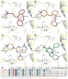
The binding pose of doxepin (magenta carbon atoms, panel A) in the H1R structure (PDB ID 3RZE) and the predicted binding poses of the novel fragment-like H1R ligands identified by prospective structure-based virtual screening: 3 (orange, B), 4 (gold, C) and 5 (green, D). The IFPs corresponding to the compounds in the displayed pose are partially presented (E). Parts of the backbone of transmembrane (TM) helices 3 4, 5, 6 and 7 are represented by transparent light yellow ribbons. Important binding residues are depicted as ball-and-sticks with grey carbon atoms. Oxygen, nitrogen, and hydrogen atoms are coloured red, blue and cyan, respectively. H-bonds described in the text are depicted by black dots. The IFP bit strings of the docking poses of the hits 3–5 (B–D) are compared to the reference IFP of doxepin 1 (A) in panel E, encoding different interaction types with each residue in the binding site. For reasons of clarity, the bit strings of only 6 residues (out of 33) are shown as an example.
Figure 3
Figure 3
Radioligand displacement by (A, B) and functional effects of (C) cpds 19. A, B: Displacement curves of [3H]-mepyramine in HEK293T cells transiently transfected with hH1R (n=3, each performed in triplicate). C: Inhibition of histamine-stimulated inositol phosphate accumulation assay in HEK293T cells transiently transfected with hH1R by cpds 1–9 (n=2, each performed in triplicate).
Figure 4
Figure 4
Hit rate and size of hits identified in prospective structure-based virtual screening studies against GPCR crystal structures and homology models. The bars shown for the heavy atom count indicate the minimum and maximum heavy atom count for all hits of each SBVS. The labels indicate the screening on the following receptors adenosine alpha-2a receptor (A2A_121 and A2A_222), adrenergic alpha-1a receptor (ADA1A10), adrenergic beta-2 receptor (ADRB220), complement component 3a receptor 1 (C3A19), C-C chemokine receptor type 5 (CCR514), cannabinoid receptor 2 (CNR213), dopamine receptor D3 (DRD38), free fatty acid receptor 1 (FFAR118), formyl peptide receptor 1 (FPR111), histamine receptor H1, histamine receptor H4 (H417), melanin-concentrating hormone receptor 1 (MCH1_115 and MCH1_29), neurokinin 1 receptor (NK1R12) and transferrin receptor protein 1 (TRFR116). The maximum heavy atom count of FPR1 (41) is not shown for clarity purposes. Only hits for which: i) binding affinity (Ki ≤ 15 μM) or potency (EC50 ≤ 15 μM) was experimentally determined; and ii) for which a molecular structure was reported are included in the analysis.
Figure 5
Figure 5
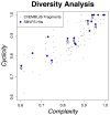
SCA plot showing the distribution of experimentally validated structure-based virtual screening hits of H1R (blue dots) in the chemical space covered by previously fragment-like H1R ligands in the ChEMBLdb. The positions of all sub-micromolar affinity hits are indicated by the numbers (see Table 2).

References

    1. Lagerstrom MC, Schioth HB. Structural diversity of G protein-coupled receptors and significance for drug discovery. Nature reviews Drug discovery. 2008;7:339–357. - PubMed
    1. Rosenbaum DM, Rasmussen SG, Kobilka BK. The structure and function of G-protein- coupled receptors. Nature. 2009;459:356–363. - PMC - PubMed
    1. Topiol S, Sabio M. X-ray structure breakthroughs in the GPCR transmembrane region. Biochemical pharmacology. 2009;78:11–20. - PubMed
    1. Congreve M, Langmead CJ, Mason JS, Marshall FH. Progress in structure based drug design for g protein-coupled receptors. Journal of medicinal chemistry. 2011;54:4283–4311. - PMC - PubMed
    1. de Graaf C, Rognan D. Customizing G Protein-coupled receptor models for structure-based virtual screening. Current pharmaceutical design. 2009;15:4026–4048. - PubMed

Publication types