. 2012 Jan;120(2):302-13.
doi: 10.1111/j.1471-4159.2011.07543.x. Epub 2011 Dec 2.

Atp8a1 deficiency is associated with phosphatidylserine externalization in hippocampus and delayed hippocampus-dependent learning

Affiliations

Atp8a1 deficiency is associated with phosphatidylserine externalization in hippocampus and delayed hippocampus-dependent learning

Kelly Levano et al. J Neurochem. 2012 Jan.

Abstract

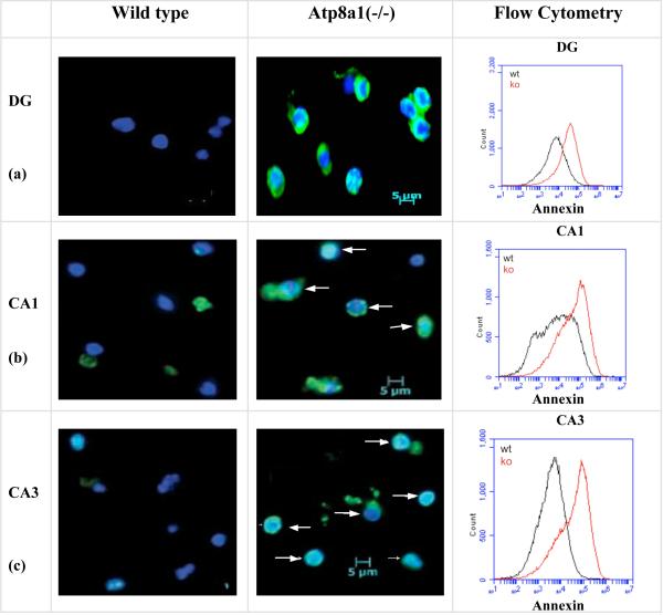
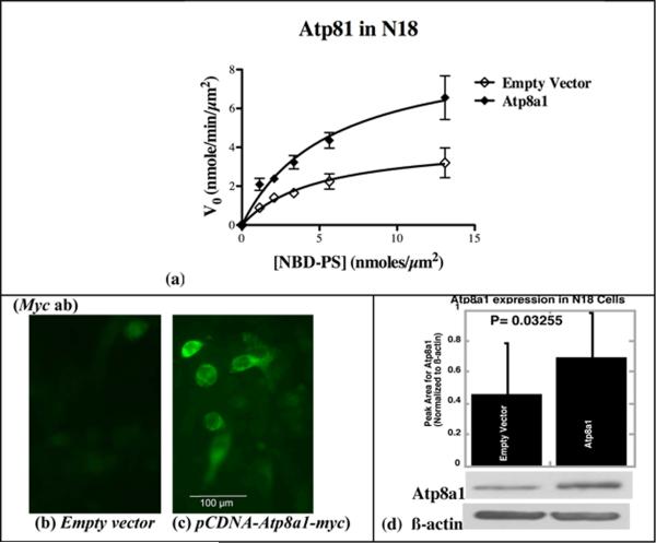
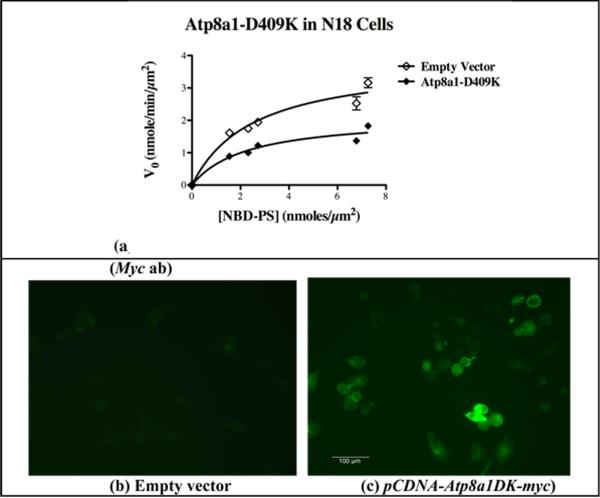
The molecule responsible for the enzyme activity plasma membrane (PM) aminophospholipid translocase (APLT), which catalyzes phosphatidylserine (PS) translocation from the outer to the inner leaflet of the plasma membrane, is unknown in mammals. A Caenorhabditis elegans study has shown that ablation of transbilayer amphipath transporter-1 (TAT-1), which is an ortholog of a mammalian P-type ATPase, Atp8a1, causes PS externalization in the germ cells. We demonstrate here that the hippocampal cells of the dentate gyrus, and Cornu Ammonis (CA1, CA3) in mice lacking Atp8a1 exhibit a dramatic increase in PS externalization. Although their hippocampi showed no abnormal morphology or heightened apoptosis, these mice displayed increased activity and a marked deficiency in hippocampus-dependent learning, but no hyper-anxiety. Such observations indicate that Atp8a1 plays a crucial role in PM-APLT activity in the neuronal cells. In corroboration, ectopic expression of Atp8a1 but not its close homolog, Atp8a2, caused an increase in the population (V(max) ) of PM-APLT without any change in its signature parameter K(m) in the neuronal N18 cells. Conversely, expression of a P-type phosphorylation-site mutant of Atp8a1 (Atp8a1*) caused a decrease in V(max) of PM-APLT without significantly altering its K(m) . The Atp8a1*-expressing N18 cells also exhibited PS externalization without apoptosis. Together, our data strongly indicate that Atp8a1 plays a central role in the PM-APLT activity of some mammalian cells, such as the neuronal N18 and hippocampal cells.


Figures

Fig. 1
Fig. 1. Genotyping of Atp8a1(-/-) mice and morphological analysis of wild type and Atp8a1(-/-) brains
(a) In genotyping analysis, the mutant allele yielded a 437-bp PCR product, whereas wild type allele produced a 253-bp product. (b) and (c) Immunohistochemistry using Ab-B confirmed the expression of Atp8a1 in the whole brain of wild type mice and its absence in the Atp8a1(-/-) mice. (d) and (e) H&E staining showing no morphological aberrance in wild type or Atp8a(-/-) mouse brains. (f) and (g) HOECHST33342 staining revealed clearly visible nucleoli in wild type and Atp8a1(-/-) mice (enlarged view of one arm of the DG layer has been presented here).
Fig. 2
Fig. 2. Spontaneous PS-externalization in the hippocampus of the Atp8a1(-/-) but not the wild type mice
(a-c) Alexafluor488-Annexin V (green) and HOECHST33342 (blue) staining of cells from the neuronal layers in DG, CA1, and CA3 of control and Atp8a1(-/-) mice. Merged images of annexin V(+) cells were observed in the cells from the Atp8a1(-/-) mice. White arrows show non-apoptotic but annexin V(+) cells. (Flow Cytometry, Right panel) From three experiments, the increases in mean fluorescence were 3.6-fold (DG), 3.9-fold (CA1), and 10.7-fold (CA3) in the Atp8a1(-/-) samples (red traces).
Fig. 3
Fig. 3. The Atp8a1(-/-) mice show marked deficiency in Morris Water Maze (MWM) and increased activity in Open Field (OF) and Elevated-Plus Maze (EPM)
(a) MWM: On each day after day 1, there was a significant difference between wild type and mutant mice (p < 0.0001) (six mice/group). (b) OF: the Atp8a1(-/-) mice displayed significantly increased mobility but an insignificant difference from wild type in center entry or time spent in the center zone. (c & d) EPM: The difference between Atp8a1(-/-) and wild type was significant for Open Arm Entry and Open Arm Time. All differences in (d) were significant. Max Speed: p = 0.022; Line Crossing: p = 0.018; Time Mobile: p = 0.014; Time Immobile: p = 0.025. Data were analyzed using two-tailed unpaired t-test with unequal variance.
Fig. 4
Fig. 4. Transient Expression of Atp8a1 in the mouse neuroblastoma cell line N18 causes an increase in Vmax without any change in Km
(a) Kinetic parameters obtained with GraphPad Prism from substrate saturation curves indicate a significant increase (1.7-fold) in Vmax of PM-APLT without any change in Km in N18 cells expressing ectopic Atp8a1 (see Table I). (b and c) Immunostaining of the pCDNA-Atp8a1-myc transfected N18 cells with a Myc antibody shows plasma membrane localization of Atp8a1-Myc (Scale: 100 μm). (d) Immunoblot analysis of enriched membrane fractions using Ab-B shows a 1.7-fold increase in Atp8a1 expression (Atp8a1 bands were normalized to β-actin, from three independent experiments and analyzed using paired t-test). (Also see Figures S2, S3 and S5, Table S1, and Text S1).
Fig. 5
Fig. 5. Expression of the Atp8a1(DK) mutant causes an inhibition of Vmax without any significant change in Km for the PM-APLT activity in the N18 cells
(a) Kinetic parameters obtained with GraphPad Prism from substrate saturation curves for PM-APLT activity indicate a significant decrease in Vmax in N18 cells expressing Atp8a1(DK) without any alteration in the value of Km (see Table II). (b and c) Immunostaining of the pCDNA-Atp8a1(DK)-myc transfected N18 cells with a Myc antibody showed plasma membrane localization of Atp8a1(DK)-Myc. (Scale: 100 μm).
Fig. 6
Fig. 6. Transient expression of Atp8a1 mutants causes PS externalization without apoptosis
(a) PS externalization (annexin V staining) occurs in the N18 cells expressing the DK mutant (or DE; Fig. S6) but not wild type Atp8a1. Co-expressed AsRed2 (red) marks the transfected cells (shown by white arrows). (Scale: 100 μm). (b) Merged (enlarged) images showing localization of annexin V staining in the AsRed2-expressing transfected cells. (c) Graphical view of the increase in annexin V(+) cells (normalized to AsRed2(+) cell number) expressing the DE or DK mutants. (also see Fig. S6). (d) Transfection with pCDNA6-Myc (vector) or pCDNA6-Atp8a1(DK)-Myc yields no apoptotic cells after 24 h, but curcumin treatment yields dark, round apoptotic N18 cells (Scale: 200 μm). (e) The curcumin-treated N18 cells but not the Atp8a1(DK) expressing cells show a dramatic increase in caspase-3/7 activity (p < 0.0001 versus all other sets). Statistical analysis was performed using ANOVA.

References

    1. Adayev T, Estephan R, Meserole S, Mazza B, Yurkow EJ, Banerjee P. Externalization of phosphatidylserine may not be an early signal of apoptosis in neuronal cells, but only the phosphatidylserine-displaying apoptotic cells are phagocytosed by microglia. J. Neurochem. 1998;71:1854–1864. - PubMed
    1. Adayev T, Ray I, Sondhi R, Sobocki T, Banerjee P. The G protein-coupled 5-HT1A receptor causes suppression of caspase-3 through MAPK and protein kinase Cα. Biochim. Biophys. Acta. 2003;1640:85–96. - PubMed
    1. Arms K, Camp P. Biology. Harcourt Brace College Publishers; New York: 1995.
    1. Bratton DL, Hensen PM. Apoptotic Cell Recognition: Will the Real Phosphatidylserine Receptor(s) Please Stand up? Current Biology. 2007;18:R76–R79. - PubMed
    1. Bratton DL, Fadok VA, Richter DA, Kailey JM, Guthrie LA, Henson PM. Appearance of phosphatidylserine on apoptotic cells requires calcium-mediated non-specific flip-flop and is enhanced by loss of the aminophospholipid translocase. J. Biol. Chem. 1997;272:26159–26165. - PubMed

Publication types

MeSH terms