. 2011 Nov 22;50(46):10126-35.
doi: 10.1021/bi201197p. Epub 2011 Oct 31.

Kinetic analysis of the unique error signature of human DNA polymerase ν

Affiliations

Kinetic analysis of the unique error signature of human DNA polymerase ν

Mercedes E Arana et al. Biochemistry. .

Abstract

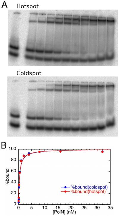
The fidelity of DNA synthesis by A-family DNA polymerases ranges from very accurate for bacterial, bacteriophage, and mitochondrial family members to very low for certain eukaryotic homologues. The latter include DNA polymerase ν (Pol ν) which, among all A-family polymerases, is uniquely prone to misincorporating dTTP opposite template G in a highly sequence-dependent manner. Here we present a kinetic analysis of this unusual error specificity, in four different sequence contexts and in comparison to Pol ν's more accurate A-family homologue, the Klenow fragment of Escherichia coli DNA polymerase I. The kinetic data strongly correlate with rates of stable misincorporation during gap-filling DNA synthesis. The lower fidelity of Pol ν compared to that of Klenow fragment can be attributed primarily to a much lower catalytic efficiency for correct dNTP incorporation, whereas both enzymes have similar kinetic parameters for G-dTTP misinsertion. The major contributor to sequence-dependent differences in Pol ν error rates is the reaction rate, k(pol). In the sequence context where fidelity is highest, k(pol) for correct G-dCTP incorporation by Pol ν is ~15-fold faster than k(pol) for G-dTTP misinsertion. However, in sequence contexts where the error rate is higher, k(pol) is the same for both correct and mismatched dNTPs, implying that the transition state does not provide additional discrimination against misinsertion. The results suggest that Pol ν may be fine-tuned to function when high enzyme activity is not a priority and may even be disadvantageous and that the relaxed active-site specificity toward the G-dTTP mispair may be associated with its cellular function(s).


Figures

Figure 1
Figure 1
Spectrum of errors generated by human Pol ν-77 at pH 7.5. The 407 template nucleotides within the single-strand gap of the M13mp2 substrate are shown as 5 lines of the template sequence, bracketed by 5 base pairs at either end to indicate the boundaries of the gap. +1 represents the first transcribed nucleotide of the lacZα-complementation region. Letters above the target sequence indicate base substitutions made by Pol ν-77. Single-base deletions are represented by open triangles below the target sequence whereas single-base additions are depicted by closed inverted triangles above the target sequence. Red characters represent phenotypically detectable changes in the gap region while black characters represent phenotypically undetectable changes found in association with detectable changes. Also depicted in the figure are the error rates at positions: 169, 165, 151, 149, 148, 145, 141, 89 and 88.
Figure 2
Figure 2
Kinetics of Pol ν-77. Panel A shows the kinetics of incorporation of dATP into the 13/19mer-T substrate (Table 1). The reactions contained 5 nM DNA primer termini, 100 μM dATP and the indicated concentrations of Pol ν-77 (measured as total protein). Because the Pol ν-77 prep was not fully active, all three concentrations of Pol ν-77 showed burst kinetics, with the burst amplitude providing a measure of the concentration of active enzyme. A plot of burst amplitude against the concentration of Pol ν-77 (Panel B) had a slope of 0.23, indicating that this Pol ν-77 preparation was 23% active. Panel C shows dTTP misinsertion by Pol ν-77 opposite template G within the G164 hotspot sequence, measured under burst conditions at a series of dTTP concentrations. The burst rate of G-dTTP misinsertion was plotted as a function of dTTP concentration and fitted to a hyperbolic equation, giving Kd(dTTP) = 35 μM, and kpol = 0.076 s−1.
Figure 3
Figure 3
Gel mobility shift experiment comparing the binding of Pol ν-77 to a T-G mismatched DNA terminus in the context of the G(165) hotspot or the G(−66) coldspot sequence. In panel A, the labeled DNA was present at 0.025 nM; the leftmost lane of each gel shows the DNA in the absence of added protein, with excess primer strand having a faster mobility than the annealed duplex. Binding of the duplex is seen in the presence of Pol ν-77 at concentrations (from left to right) of 0.05, 0.1, 0.25, 0.5, 1, 2, 4, 8, 16, and 32 nM. A plot of bound DNA against Pol ν-77 concentration (Panel B), fitted to a quadratic equation (56), gave KD values of 0.38 and 0.40 nM for the hotspot and coldspot sequences respectively. Because only 23% of the Pol ν-77 was active, the true KD is 0.1 nM.
Figure 4
Figure 4
Regions of the A-family DNA polymerase structure where Pol ν shows interesting differences from the classical A-family conserved motifs. Panel A shows the structure of the polymerase domain of the ternary (Pol-DNA-dNTP) complex of Klentaq (3KTQ, ref. (54)). The protein structure is shown in grey, with residues 501-523 at the tip of the thumb subdomain omitted to avoid obscuring the active site region. The DNA duplex is shown in blue, with the template strand darker, and the incoming dNTP in cyan with CPK coloring. Protein sequence motifs discussed in the text are colored: the N-terminus of the N-helix (including the conserved histidine side-chain) in red, the O-helix in yellow, and the N-terminal portion of Motif 6, within the Q-helix, in green. Panel B shows the alignment of the corresponding portions of sequence from human Pol ν, compared with Klenow fragment and Klentaq, representing the classical A-family DNA polymerases. The motif designations are from Patel et al. (23). Residues highly conserved in the alignment of both classical and Pol ν-type A-family DNA polymerases (51) are highlighted with the colors of the corresponding motifs in Panel A.

Similar articles

Cited by

References

    1. Bebenek K, Kunkel TA. Functions of DNA polymerases. Adv. Protein Chem. 2004;69:137–165. - PubMed
    1. Marini F, Kim N, Schuffert A, Wood RD. POLN, a nuclear PolA family DNA polymerase homologous to the DNA cross-link sensitivity protein Mus308. J. Biol. Chem. 2003;278:32014–32019. - PubMed
    1. Pang M, McConnell M, Fisher PA. The Drosophila mus 308 gene product, implicated in tolerance of DNA interstrand crosslinks, is a nuclear protein found in both ovaries and embryos. DNA Repair (Amst) 2005;4:971–982. - PubMed
    1. Boyd JB, Sakaguchi K, Harris PV. mus308 mutants of Drosophila exhibit hypersensitivity to DNA cross-linking agents and are defective in a deoxyribonuclease. Genetics. 1990;125:813–819. - PMC - PubMed
    1. Harris PV, Mazina OM, Leonhardt EA, Case RB, Boyd JB, Burtis KC. Molecular cloning of Drosophila mus308, a gene involved in DNA cross-link repair with homology to prokaryotic DNA polymerase I genes. Mol. Cell Biol. 1996;16:5764–5771. - PMC - PubMed

Publication types