. 2012 Oct;15(9):1287-94.
doi: 10.1017/S146114571100143X. Epub 2011 Oct 19.

Distinct prefrontal cortical regions negatively regulate evoked activity in nucleus accumbens subregions

Affiliations

Distinct prefrontal cortical regions negatively regulate evoked activity in nucleus accumbens subregions

Amber Asher et al. Int J Neuropsychopharmacol. 2012 Oct.

Abstract

Deficits in prefrontal cortical activity are consistent observations in a number of psychiatric diseases with two major regions consistently implicated being the medial prefrontal cortex (mPFC) and orbitofrontal cortex (OFC), regions that carry out independent, but complementary forms of cognitive processing in changing environmental conditions. Information from the prefrontal cortex is integrated in the nucleus accumbens (NAc) to guide goal-directed behaviour. Anatomical studies have demonstrated that distinct prefrontal cortical regions provide an overlapping but distinct innervation of NAc subregions; however, how information from these distinct regions regulates NAc output has not been conclusively demonstrated. Here we demonstrate that, while neurons receiving convergent glutamatergic inputs from the mPFC and OFC have a synergistic effect on single-spike firing, medium spiny neurons that receive a monosynaptic input from only one region are actually inhibited by activation of the complementary region. Therefore, the mPFC and OFC negatively regulate evoked activity within the lateral and medial regions of the NAc, respectively, and exist in a state of balance with respect to their influence on information processing within ventral striatal circuits.


Figures
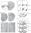
Figure 1
Figure 1
Representative brain sections demonstrating the localization of stimulating electrode placements (arrows) within the mPFC and OFC (A) and recording electrode locations within the lateral (B expanded in D) and medial (C expanded in E) NAc. The locations of each identified recording electrode (f: lateral octagons - medial squares), stimulating electrode (G: star) or infusion site (g: circle) are depicted with reference to a stereotaxic atlas adapted from Paxinos and Watson (Paxinos and Watson, 1986).
Figure 2
Figure 2
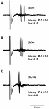
A subset of medium spiny neurons in the NAc receive converging inputs from both the mPFC and OFC. In medium spiny neurons that responded to electrical stimulation of both the OFC (A) and mPFC (B), the simultaneous activation of both PFC regions induced an augmented response than that observed with each input alone (C). Data in parentheses reflects percent spike probability.
Figure 3
Figure 3
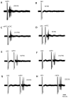
A subset of medium spiny neurons in the NAc are inhibited by prefrontal cortical activation. A representative neuron in the medial NAc that is excited by electrical activation the mPFC (A), but not OFC (B). The simultaneous activation of both afferents (C) did not appear to alter spike probability compared to mPFC activation alone. Each trace from D-H represents an increasing interstimulus interval of 10ms. Interestingly, stimulation of the OFC prior to the mPFC significantly attenuated the response to mPFC stimulation when separated by 10-30ms. Data in parentheses reflects percent spike probability.
Figure 4
Figure 4
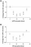
Group data demonstrating the negative regulation of evoked NAc activity by PFC subregions. Neurons recorded in the lateral NAc (A) were primarily activated by the OFC and in these neurons a preceding input from the mPFC significantly attenuates OFC-evoked responses (* represents significant difference from OFC activation alone: p<0.05, 1-way ANOVA; n = 12). Similarly, in medial NAc neurons activated by mPFC stimulation (B), the preceding activation of OFC afferents significantly inhibited evoked activity (* represents significant difference from mPFC activation alone: p<0.05, 1-way ANOVA; n = 13).
Figure 5
Figure 5
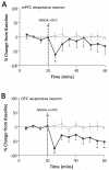
Chemical activation of PFC subregions negatively regulates evoked activity in NAc subregions. NMDA activation of the OFC significantly attenuated mPFC-evoked activity in the medial NAc (A: * represents significant difference from vehicle: p<0.05, 2-way ANOVA; n = 5). Similarly, NMDA activation of the mPFC significantly inhibited the OFC-evoked spike firing in lateral NAc neurons (B: * represents significant difference from vehicle: p<0.05, 2-way ANOVA; n = 5).

Similar articles

Cited by

References

    1. Abi-Dargham A, Mawlawi O, Lombardo I, Gil R, et al. Prefrontal Dopamine D1 Receptors and Working Memory in Schizophrenia. Journal of Neuroscience. 2002;22(9):3708–3719. - PMC - PubMed
    1. Berendse HW, Galis-de Graaf Y, Groenewegen HJ. Topographical organization and relationship with ventral striatal compartments of prefrontal corticostriatal projections in the rat. Journal of Comparative Neurology. 1992;316(3):314–347. - PubMed
    1. Birrell JM, Brown VJ. Medial frontal cortex mediates perceptual attentional set shifting in the rat. Journal of Neuroscience. 2000;20(11):4320–4324. - PMC - PubMed
    1. Brog JS, Salyapongse A, Deutch AY, Zahm DS. The patterns of afferent innervation of the core and shell in the “accumbens” part of the rat ventral striatum: immunohistochemical detection of retrogradely transported fluoro-gold. Journal of Comparative Neurology. 1993;338(2):255–278. - PubMed
    1. Chakirova G, Welch KA, Moorhead TWJ, Stanfield AC, et al. Orbitofrontal morphology in people at high risk of developing schizophrenia. European Psychiatry. 2010;25(6):366–372. - PubMed

Publication types

MeSH terms