. 2012 Jan 15;361(2):191-207.
doi: 10.1016/j.ydbio.2011.09.031. Epub 2011 Oct 10.

Differential requirements for Myocyte Enhancer Factor-2 during adult myogenesis in Drosophila

Affiliations

Differential requirements for Myocyte Enhancer Factor-2 during adult myogenesis in Drosophila

Anton L Bryantsev et al. Dev Biol. .

Abstract

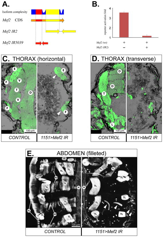
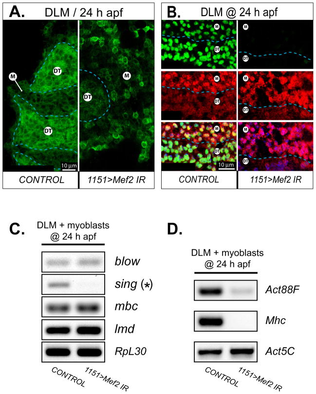
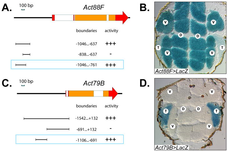
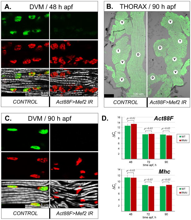
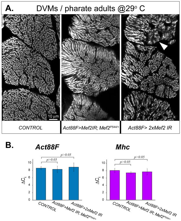
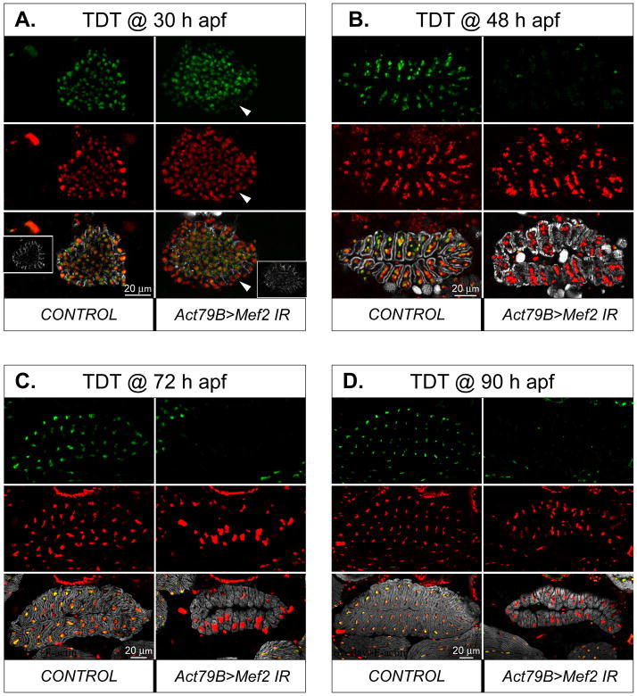
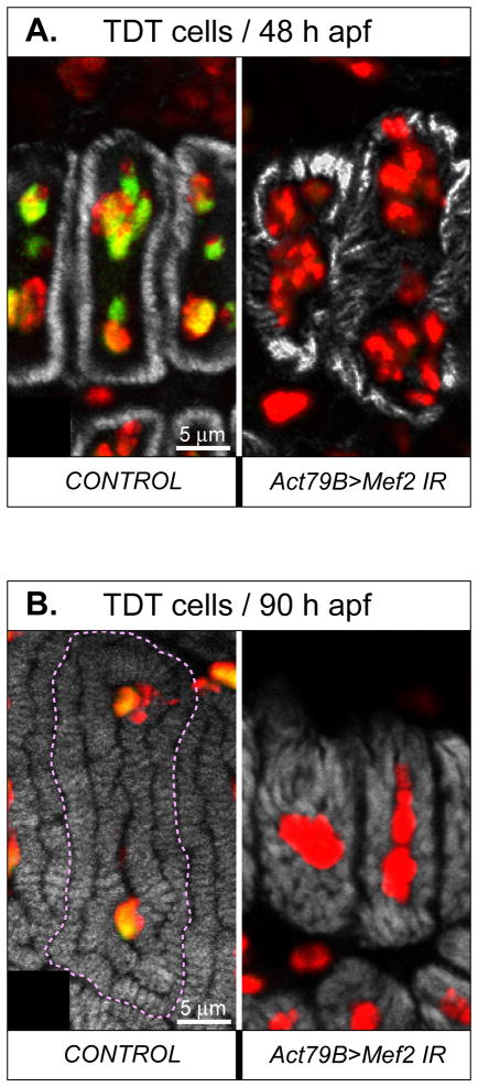
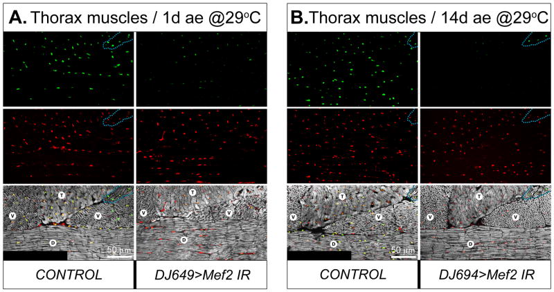
Identifying the genetic program that leads to formation of functionally and morphologically distinct muscle fibers is one of the major challenges in developmental biology. In Drosophila, the Myocyte Enhancer Factor-2 (MEF2) transcription factor is important for all types of embryonic muscle differentiation. In this study we investigated the role of MEF2 at different stages of adult skeletal muscle formation, where a diverse group of specialized muscles arises. Through stage- and tissue-specific expression of Mef2 RNAi constructs, we demonstrate that MEF2 is critical at the early stages of adult myoblast fusion: mutant myoblasts are attracted normally to their founder cell targets, but are unable to fuse to form myotubes. Interestingly, ablation of Mef2 expression at later stages of development showed MEF2 to be more dispensable for structural gene expression: after myoblast fusion, Mef2 knockdown did not interrupt expression of major structural gene transcripts, and myofibrils were formed. However, the MEF2-depleted fibers showed impaired integrity and a lack of fibrillar organization. When Mef2 RNAi was induced in muscles following eclosion, we found no adverse effects of attenuating Mef2 function. We conclude that in the context of adult myogenesis, MEF2 remains an essential factor, participating in control of myoblast fusion, and myofibrillogenesis in developing myotubes. However, MEF2 does not show a major requirement in the maintenance of muscle structural gene expression. Our findings point to the importance of a diversity of regulatory factors that are required for the formation and function of the distinct muscle fibers found in animals.


Figures

Fig. 1
Fig. 1. Structural and functional properties of the inverted repeat (IR) RNAi constructs
A: Mapping the sequences used in two different IR constructs to the coding DNA sequence (CDS) of Mef2. Isoform complexity depicts the usage of nucleotide sequence in Mef2 annotated transcripts, with the highest plotted value corresponding to usage in all transcripts. B: Mef2 silencing ability of the Mef2 IR2 construct assayed in cell culture co-transfection assays. Reporter activation was determined as fold β-galactosidase accumulation, over the basal β-galactosidase expression level when the activating Mef2-expressing plasmid was omitted. Expression of β-galactosidase from the reporter construct was controlled by a MEF2-resposive enhancer/promoter of the Act57B gene. C, D, E: Comparative analysis of gross muscle morphology between control (on the left of each panel) and RNAi (on the right of each panel) pharate unhatched adults at 90 h APF on indicated planes of thorax sections (C, D) and abdominal fillets (E), stained for F-actin. The RNAi was activated by the 1151-Gal4 driver in adult myoblast precursors. The UAS-Mef2 IR constructs were: IR2 (C) and IR5039 (D, E). Arrowheads point to escaper muscles that did form; white box indicates unusual mini-muscles. E – extracoxal depressor of the trochanter, F – direct flight muscles, T – TDT, D – DLM, V – DVM, L – leg muscles, TL – dorsal internal oblique muscles, which are temporary muscles of larval origin, AD – abdominal dorsal muscles, AL – abdominal lateral muscles, H – cardiac muscles of the dorsal vessel.
Fig. 2
Fig. 2. Induction of RNAi in adult muscle precursors severely affects myogenesis by preventing myoblasts from fusing into myofiber syncytia
A: Transverse, hematoxylin/eosin stained section of the thorax of control and RNAi pupae at 24 h APF, showing DLM templates (DT) surrounded with swarming myoblasts (M). Note that the number of DLM templates in RNAi-induced animal is not doubled, and remains at three. B: Transversely cut developing DVM fibers on horizontal sections of the thorax of control and RNAi pupae at 24 h APF. Control has a myofiber core for each fiber (C), which is absent in the RNAi animal. C, D: horizontal sections of the thorax of control and RNAi pupae at 30 h (C) and 48 h (D) APF, respectively. In control animals, diverse types of muscles have been formed, while in RNAi animals there are only myoblasts which become sparse at 48 h APF. The RNAi was induced with the 1151-Gal4 driver activating UAS-Mef2 IR5039. DT – DLM template, M – myoblasts, C – fusion core, D – DLM, V – DVM, T – TDT.
Fig. 3
Fig. 3. Mef2 knockdown in adult myoblasts compromises myoblast behavior at fusion centers and prevents expression of fusion and muscle structural genes
A: Transverse sections across developing DLM fibers at 24 h APF are shown for control and RNAi (1151>Mef2 IR5039) conditions. The signal is recorded from membrane-associated mCD8:GFP protein driven by the same 1151-Gal4 driver as used for RNAi induction. Individual myoblasts make tight formations around the outlined DLM fusion templates (DT) in the control animal, but only loosely aggregate around DT in the RNAi animal. Note, that mCD8 also labels the nuclear envelopes within the fusion templates. B: A boundary area between a DT and unfused myoblasts (M) at 24 h APF, demonstrating a substantial decrease in MEF2 immunofluorescence (green) in both myoblasts and fusion templates, upon induction of Mef2 RNAi with the 1151-Gal4 driver. Myoblasts are traced with the twi-LacZ transgene whose expression is detected by immunofluorescent staining for β-galactosidase (red). The twi-lacZ activity is subdued in DT, identifying its border with unfused myoblasts (dashed line). The overlaid images at the bottom of the panel are additionally counter-stained for nuclei (blue). C: Expression analysis of several fusion-critical genes in samples obtained at the sites of developing DLMs at 24 h APF, from control and RNAi (1151>Mef2 IR5039) animals. Fusion genes are indicated with red font. The asterisk marks the sing gene, that shows a significantly reduced expression in response to Mef2 knockdown. The transcription factor lmd is used as a genetic marker of myoblasts; and the ribosomal protein gene, RpL30, is used as a general marker for RT-PCR loading control. D: Activation of muscle structural genes using RT-PCR and detecting expression of the pan-muscular Mhc, and the IFM-specific Act88F structural genes in the same samples as in C. The housekeeping actin, Act5C, is used as loading control.
Fig. 4
Fig. 4. Characterization of the enhancer regions used in post-fusion-activated Gal4 drivers
A, C: The structure of Act88F (A) and Act79B (C) genes. Red and orange rectangles indicate non-coding and coding exons, respectively; white boxes represent introns. The cloned fragments used for enhancer activity analysis, their precise boundaries related to the transcriptional initiation site, and in vivo activities are indicated below each gene scheme. All drawings are to scale and oriented to the respective gene schemes. The selected enhancers, used in Gal4 drivers, are framed. B, D: Expression of Act88F-Gal4 (B) and Act79B-Gal4 (D) drivers, as revealed by activation of a UAS-LacZ construct. β-galactosidase enzymatic activity was detected in situ on horizontal thorax sections using X-gal. T – TDT, D – DLM, V – DVM.
Fig. 5
Fig. 5. Morphological and molecular effects of Mef2 silencing in post-fused IFM fibers
A: Expression of MEF2 (green) in the nuclei (red), as well as nascent myofibrils (gray), in longitudinally sectioned DVM fibers shortly after completion of myoblast fusion in control and Act88F-Gal4-induced RNAi (Act88F>Mef2 IR5039) pupae at 48 h APF. B: General muscle localization in the thorax of control and RNAi pharate adults at 90 h APF, visualized by phalloidin staining (green). The DLM fibers (D) in RNAi animals (Act88F>Mef2 IR5039) are collapsed. The positions of other major thoracic muscles, DVMs (V) and TDT (T), remain unchanged. C: Longitudinally sectioned DVMs, stained for MEF2 (green), nuclei (red), and myofibrils (gray) in control and RNAi pharate adults (Act88F>Mef2 IR5039) at 90 h APF. Lack of MEF2 results in a wavy arrangement of myofibrils, although their structure is similar to control. D: Comparative analysis of expression of Act88F (upper panel) and Mhc (lower panel) between control (green bars, WT) and Act88F>Mef2 IR5039 (red bars, RNAi) samples, obtained from IFMs (combined DVM and DLM fibers) at 48 h, 72 h, and 90 h APF. Realtime qRT-PCR data are shown as the lag in detection (ΔCt) between transcripts of interest, and 18S and 28S rRNA, used as internal references (details are in Material and Methods) ± s.d. Student’s t-test p value > 0.05 indicates statistically non-significant differences. Note that the expression of Act88F lags slightly in the RNAi samples at the 48 h APF time-point, but not at any other stages. Mhc transcript levels are not significantly different between control and knockdown samples.
Fig. 6
Fig. 6. Enhanced knockdown of Mef2 in post-fused DVMs worsens myofibril morphology, but does not affect expression of two major structural genes
A: Phalloidin-stained transverse sections through posterior DVM fibers, from pharate adults in which Mef2 RNAi was either not induced (CONTROL), induced with a single IR5039 construct in an heterozygous Mef2P544/+ null allele background (Act88F>Mef2 IR; Mef2P544/+), or induced with both IR2 and IR5039 constructs (Act88F>2xMef2 IR). RNAi was initiated and maintained via the IFM-specific Act88F-Gal4 driver. To reach maximum RNAi effectiveness, pupae were allowed to develop at 29°C. Note that under these exacerbated conditions, myofibril structure is significantly perturbed, showing misaligned, thinned myofibrils, occasionally lacking any structure (arrowhead). The phenotype conferred by double IRs was most severe. B: Expression analysis of Act88F and Mhc genes in combined samples of DLM and DVM fibers collected from the pharate adults described above. The qRT-PCR data, shown as detection delay (ΔCt) from internal reference rRNA transcripts, do not show statistically significant differences between all samples (t-test p value > 0.05), indicating that expression of both Act88F and Mhc does not readily respond to Mef2 knockdown, even under enhanced conditions.
Fig. 7
Fig. 7. Mef2 knockdown in developing TDTs results in moderate, but persistent, changes in muscle morphology
A-D: Transverse section through developing TDT fibers at 30 h (A), 48 h (B), 72 h (C), and 90 h [pharate adult] (D) APF in control and (1151>Mef2 IR5039) RNAi pupae. Samples are immunofluorescently stained for MEF2 (green), DNA (red), and F-actin (gray). The Mef2 silencing was induced with the Act79B-Gal4 driver and was evident by reduced MEF2 immunofluorescence, readily noticeable in the nuclei of fused fibers at 24 h APF (arrowhead). Insets in panel A show the appearance of developing muscle fibers at the periphery of the myoblast swarm, that contain increasing amount of polymerized actin; the MEF2 positive nuclei in the center belong to unfused myoblasts. Note that, at later stages, MEF2 immunofluorescence is either severely reduced (B) or not detected (C, D) in TDT, but stay at high levels in unaffected neighboring muscles. Anterior is to the left.
Fig. 8
Fig. 8. Mef2 knockdown in the TDT does not prevent myofibrillogenesis, but affects organization of myofibril arrays and general myofiber morphology
A, B: Transversely cut individual TDT muscle fibers at 48 h (A) and 90 h (B) APF, in control and RNAi (1151>Mef2 IR5039) pupae. The superimposed images of three fluorescent channels, corresponding to MEF2 (green), DNA (red), and F-acting (grey) are shown. The yellow color indicates an overlap between the green and the red signals. Note that nuclei of RNAi fibers contain only the red signal and are significantly disorganized. Fibrillar organization is disturbed in RNAi fibers at both time points, but the sizes of individual myofibrils have increased between 48 h and 90 h APF.
Fig. 9
Fig. 9. Mef2 knockdown in post-developed IFMs does not compromise muscle morphology
A, B: Major thoracic muscles of control and RNAi (DJ694>Mef2 IR2) males at 1 day ae (ae) (A), or 14 days ae (B), stained for MEF2 (red), DNA (green), and F-actin (grey). The flies were raised at 29°C. Note that MEF2 immunofluorescence in DLMs (D) and DVMs (V) is reduced at 1day ae, and not detected at 14 days ae in RNAi flies, while staying unchanged in control and RNAi non-affected direct flight muscles (outlined with dashed line). MEF2 immunofluorescence in the TDT (T) of RNAi flies is also reduced, but to a lesser extent than in DVMs and DLMs. Anterior is to the left.
Fig. 10
Fig. 10. RNAi knockdown of the actin gene Act88F in post-developed muscles does not alter myofibril morphology
A: Overlaid images of an enlarged area of the DLM in control and RNAi pupae at 72 h APF, stained for MEF2 (green), DNA (red), and F-actin (grey); overlaid red and green signals produce yellow. The RNAi was induced by Act88F-Gal4 driving the Act88F IR construct. In RNAi-induced DLM myofibrils are missing, demonstrating the effectiveness of the Act88F IR construct. B: Enlarged area of the DLM stained for F-actin in control and RNAi flies staged for 31 days at 29°C. The adult-specific RNAi was induced by the DJ694-Gal4 driver and the same Act88F IR construct. Unlike the situation in the developing muscles, post-developed muscles in adults do not produce a fibrillar phenotype in response to actin silencing, even after a prolonged exposure to RNAi targeting Act88F.

References

    1. Anant S, Roy S, VijayRaghavan K. Twist and Notch negatively regulate adult muscle differentiation in Drosophila. Development. 1998;125:1361–9. - PubMed
    1. Andres V, Cervera M, Mahdavi V. Determination of the consensus binding site for MEF2 expressed in muscle and brain reveals tissue-specific sequence constraints. J Biol Chem. 1995;270:23246–9. - PubMed
    1. Apitz H. pChs-Gal4, a vector for the generation of Drosophila Gal4 lines driven by identified enhancer elements. Dros Inf Serv. 2002;85:118–120.
    1. Arredondo JJ, Ferreres RM, Maroto M, Cripps RM, Marco R, Bernstein SI, Cervera M. Control of Drosophila paramyosin/miniparamyosin gene expression. Differential regulatory mechanisms for muscle-specific transcription. J Biol Chem. 2001;276:8278–87. - PubMed
    1. Atreya KB, Fernandes JJ. Founder cells regulate fiber number but not fiber formation during adult myogenesis in Drosophila. Dev Biol. 2008;321:123–40. - PubMed

Publication types

MeSH terms

Substances