. 2012 Feb;20(2):347-55.
doi: 10.1038/mt.2011.215. Epub 2011 Oct 18.

Rad51 promoter-targeted gene therapy is effective for in vivo visualization and treatment of cancer

Affiliations

Rad51 promoter-targeted gene therapy is effective for in vivo visualization and treatment of cancer

Christopher M Hine et al. Mol Ther. 2012 Feb.

Abstract

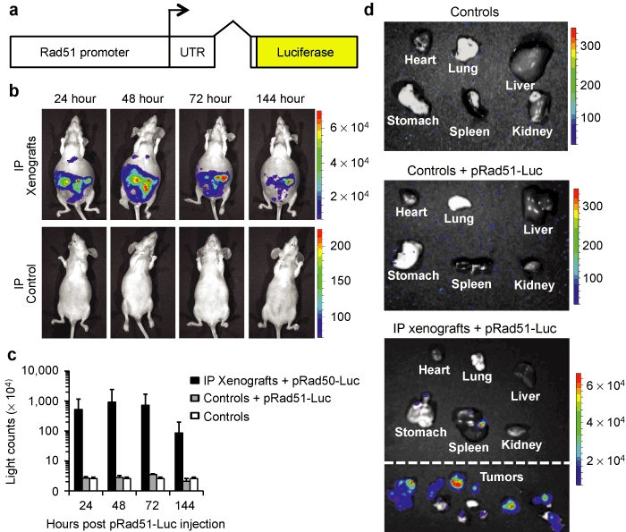
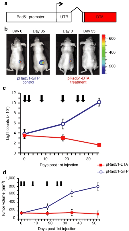
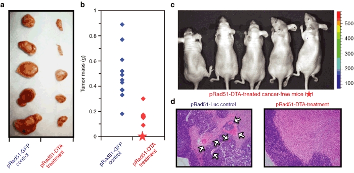
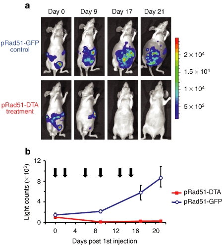
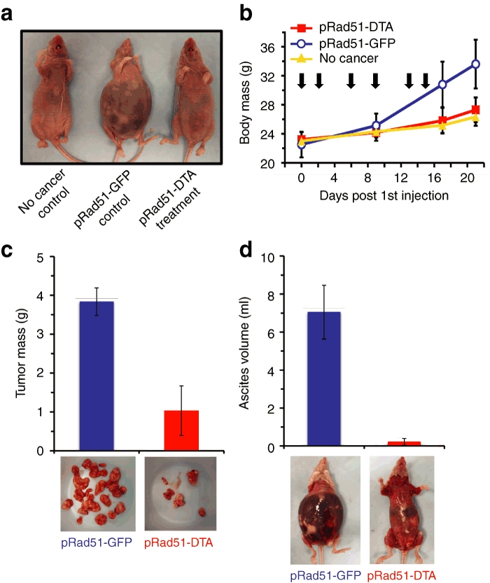
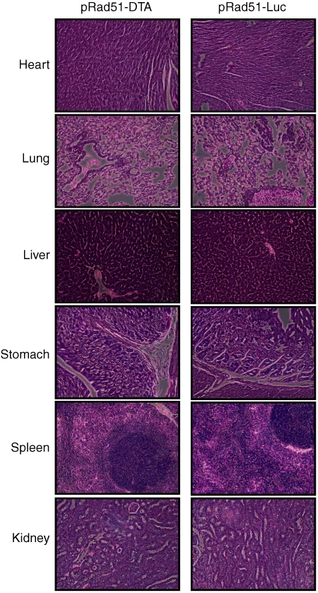
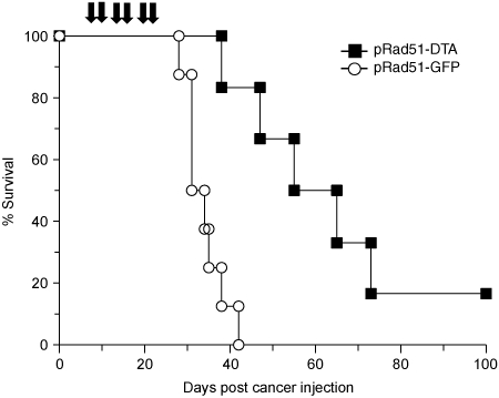
Rad51 protein is overexpressed in a wide range of human cancers. Our previous in vitro studies demonstrated that a construct comprised Rad51 promoter driving expression of the diphtheria toxin A gene (pRad51-diphtheria toxin A (DTA)) destroys a variety of human cancer cell lines, with minimal to no toxicity to normal human cells. Here we delivered Rad51 promoter-based constructs in vivo using linear polyethylenimine nanoparticles, in vivo jetPEI, to visualize and treat tumors in mice with HeLa xenografts. For tumor detection, we used pRad51-Luc, a construct containing the firefly luciferase under the Rad51 promoter, administered by intraperitoneal (IP) injection. Tumors were detected with an in vivo bioluminescent camera. All mice with cancer displayed strong bioluminescence, while mice without cancer displayed no detectable bioluminescence. Treatment with pRad51-DTA/jetPEI decreased tumor mass of subcutaneous (SC) and IP tumors by sixfold and fourfold, respectively, along with the strong reduction of malignant ascites. Fifty percent of the mice with SC tumors were cancer-free after six pRad51-DTA/jetPEI injections, and for the mice with IP tumors, mean survival time increased by 90% compared to control mice. This study demonstrates the clinical potential of pRad51-based constructs delivered by nanoparticles for the diagnostics and treatment of a wide range of cancers.


Figures

Figure 1
Figure 1
Visualization of IP xenografts using pRad51-Luc construct delivered by nanoparticles. (a) Diagram of pRad51-Luc construct, containing Rad51 promoter, including the 5' UTR, first intron, and first 13 amino acids of the Rad51 ORF fused in-frame with the gene encoding firefly luciferase. (b) Representative images of a mouse with xenografts and a control mouse injected with pRad51-Luc. Athymic nude-Foxn1nu mice were injected IP with HeLa cancer cells or kept cancer free. Three weeks later; mice were injected IP with pRad51-Luc complexed with in vivo-jetPEI nanoparticles. At indicated times post pRad51-Luc injection, tumor-bearing and control mice were imaged for in vivo bioluminescence on a CCD camera at 1-minute exposures. Color scale bar indicates fluorescence intensity in photons per second. (c) Quantification of the in vivo bioluminescence data for mice with tumors, tumor-free mice injected with pRad51-Luc, and untreated tumor-free mice (n = 4, for each group). (d) Ex vivo bioluminescent imaging of organs and tumor xenografts from control mice, control mice injected with pRad51-Luc, and mice bearing IP xenografts injected IP with pRad51-Luc. Images were taken 48 hours after injection. ORF, open reading frame.
Figure 2
Figure 2
pRad51-DTA delivered by nanoparticles selectively destroys SC cancer xenografts. (a) Diagram of pRad51-DTA construct, containing Rad51 promoter, 5' UTR, first intron, and first 13 amino acids of the Rad51 ORF fused in-frame to the gene encoding bacteria diphtheria toxin A. (b) HeLa cells stably expressing firefly luciferase were used to establish xenografts in athymic nude-Foxn1nu mice. Three weeks after xenograft inoculation, mice were given a series of six intratumoral injections of pRad51-DTA/jetPEI or pRad51-GFP/jetPEI (n = 10 for each group) over 28 days. Shown are representative images of mice bearing SC HeLa-Luc xenografts treated with pRad51-DTA or control pRad51-GFP at the start of the treatment and after six treatment injections. Images were taken at 2-second exposures using bioluminescent CCD camera. (c) Quantification of tumor load using the bioluminescent light counts. (d) Quantification of tumor volume measured by calipers. Arrows indicate the days of DNA/nanoparticle injections. ORF, open reading frame.
Figure 3
Figure 3
Treatment with pRad51-DTA nanoparticles efficiently reduces or eliminates SC HeLa-Luc cancer xenografts. (a) Tumors from control (pRad51-GFP) and pRad51-DTA-treated mice. The tumors were excised 25 days after the last therapy injection, imaged with a digital camera and then weighed. The largest tumors from the treated mice are shown, as the tumors in the remaining five mice were undetectable by calipers, and these mice were left alive to observe tumor recurrence. (b) Tumor mass of the tumors excised from the control and treated mice 25 days after the last injection. The star indicates five mice whose tumors became undetectable after therapy. (c) Five of the treated mice with undetectable tumors. A 1-minute exposure 100 days after the last pRad51-DTA/jetPEI injection reveals no detectable luminescence from HeLa-Luc cells. At 12 months after therapy, the mice did not show any tumor recurrence. (d) H&E staining of tumors excised from mice after six injections of pRad51-Luc (control) or pRad51-DTA (treatment). Arrows indicate locations of blood vessels where angiogenesis has occurred. ORF, open reading frame.
Figure 4
Figure 4
Treatment with pRad51-DTA nanoparticles reduces tumor burden in mice with IP HeLa-Luc xenografts. HeLa cells stably expressing firefly luciferase were used to establish xenografts in athymic nude-Foxn1nu mice. One week after xenograft inoculation, mice were given a series of six IP injections of pRad51-DTA/jetPEI or pRad51-GFP/jetPEI (n = 8 for each group) over 15 days. (a) Representative images of mice bearing IP HeLa-Luc xenografts treated with IP injections of pRad51-DTA or control pRad51-GFP at the start of the treatment and after the six treatment injections. Images were taken at 5-second exposures using bioluminescent CCD camera. (b) Quantification of tumor load using the bioluminescent light counts. Arrows indicate the days of DNA/nanoparticle injections.
Figure 5
Figure 5
IP injections of pRad51-DTA nanoparticles reduce tumor burden and malignant ascites in mice carrying IP xenografts. (a) Representative images of untreated tumor-free mice, and mice with IP xenografts that received the pRad51-GFP control or pRad51-DTA treatment. (b) Changes in body mass of mice receiving treatments or of the control tumor-free mice of the same age. Body mass of the cancer-free mice increases slightly because of animal growth. Body mass of the tumor-bearing mice receiving pRad51-GFP increased because of tumor growth and development of ascites. The increase in body mass in the pRad51-GFP-treated group was 2.7-fold greater than that of the increase in body mass in the pRad51-DTA group (P = 0.0003). There was no significant difference (P = 0.3998) in body mass between cancer-free mice and pRad51-DTA-treated cancer mice indicating that the treatment efficiently reduced tumor growth and ascites. Furthermore, there was no weight loss in the treated group indicating low toxicity. (c) Tumors and (d) ascites fluid were removed on day 21 from pRad51-DTA-treated (n = 4) and pRad51-GFP control (n = 4) mice. Mice treated with pRad51-DTA had 3.7-fold lower tumor mass (P = 0.0003) and 32.9-fold less ascites fluid (P > 0.0001) than pRad51-GFP control mice.
Figure 6
Figure 6
pRad51-DTA treatment does not result in observable toxicity to normal, healthy tissues. H&E staining of tissue samples taken from athymic nude-Foxn1nu mice after the administration of six IP injections of 100 µg pRad51-DTA or pRad5-Luc complexed with in vivo-jetPEI over a 2-week period.
Figure 7
Figure 7
Treatment with pRad51-DTA extends survival of mice with IP HeLa xenografts. Mice were injected IP with pRad51-DTA or the control pRad51-GFP nanoparticles. Arrows indicate the days of injection. The 50% survival point for pRad51-DTA-treated mice is extended by 90% over the control group (P = 0.0027). By day 42, all of the control mice had died (n = 8), while 80% of the pRad51-DTA-treated mice were alive (n = 6). *One pRad51-DTA-treated mouse was tumor-free after the treatment and remained tumor free until it was euthanized 9 months later.

Similar articles

Cited by

References

    1. Baumann P., and, West SC. Role of the human RAD51 protein in homologous recombination and double-stranded-break repair. Trends Biochem Sci. 1998;23:247–251. - PubMed
    1. Petermann E, Orta ML, Issaeva N, Schultz N., and, Helleday T. Hydroxyurea-stalled replication forks become progressively inactivated and require two different RAD51-mediated pathways for restart and repair. Mol Cell. 2010;37:492–502. - PMC - PubMed
    1. Hine CM, Seluanov A., and, Gorbunova V. Use of the Rad51 promoter for targeted anti-cancer therapy. Proc Natl Acad Sci USA. 2008;105:20810–20815. - PMC - PubMed
    1. Richardson C. RAD51, genomic stability, and tumorigenesis. Cancer Lett. 2005;218:127–139. - PubMed
    1. Miyagawa K. Clinical relevance of the homologous recombination machinery in cancer therapy. Cancer Sci. 2008;99:187–194. - PMC - PubMed

Publication types

MeSH terms