. 2011 Oct 18;30(22):4616-27.
doi: 10.1038/emboj.2011.377.

Structure and activity of the Cas3 HD nuclease MJ0384, an effector enzyme of the CRISPR interference

Affiliations

Structure and activity of the Cas3 HD nuclease MJ0384, an effector enzyme of the CRISPR interference

Natalia Beloglazova et al. EMBO J. .

Abstract

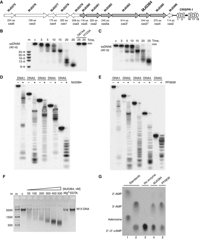
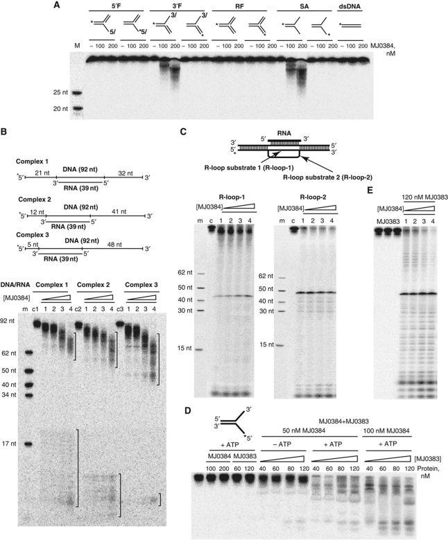
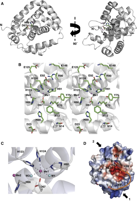
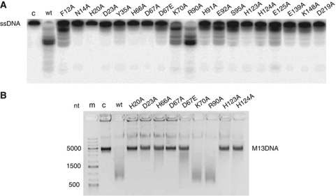
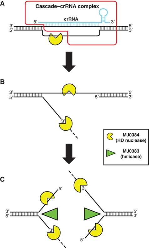
Clustered regularly interspaced short palindromic repeats (CRISPRs) and Cas proteins represent an adaptive microbial immunity system against viruses and plasmids. Cas3 proteins have been proposed to play a key role in the CRISPR mechanism through the direct cleavage of invasive DNA. Here, we show that the Cas3 HD domain protein MJ0384 from Methanocaldococcus jannaschii cleaves endonucleolytically and exonucleolytically (3'-5') single-stranded DNAs and RNAs, as well as 3'-flaps, splayed arms, and R-loops. The degradation of branched DNA substrates by MJ0384 is stimulated by the Cas3 helicase MJ0383 and ATP. The crystal structure of MJ0384 revealed the active site with two bound metal cations and together with site-directed mutagenesis suggested a catalytic mechanism. Our studies suggest that the Cas3 HD nucleases working together with the Cas3 helicases can completely degrade invasive DNAs through the combination of endo- and exonuclease activities.


Conflict of interest statement

The authors declare that they have no conflict of interest.

Figures

Figure 1
Figure 1
M. jannaschii CRISPR cluster 1 and nuclease activity of MJ0384 and PF0639. (A) Schematic representation of the M. jannaschii cas genes associated with the CRISPR cluster 1. (B, C) Time course of the linear ssDNA hydrolysis. The 5′-[32P]-labelled ssDNA (40 nt) was incubated without protein (lane c) or with wild-type and inactive MJ0384 mutant proteins (B; 200 nM) or PF0639 (C; 300 nM) for the indicated times. (D, E) Cleavage of several ssDNAs with various sequences and lengths. The 5′-[32P]-labelled ssDNAs (0.1 μM; 17–92 nt) were incubated without (−) or with MJ0384 (200 nM, 10 min) or PF0639 (300 nM, 20 min). (F) Cleavage of the circular M13mp18 ssDNA by MJ0384. The M13 ssDNA (5 nM) was incubated for 30 min without enzyme (lane c) or with various amounts of MJ0384 (50–500 nM) and the reaction products were analysed by agarose gel electrophoresis and SYBR Green staining. The last two lanes show additional controls without Mg2+ addition (−Mg2+) or in the presence of 10 mM EDTA. Lane m, DNA markers. (G) Hydrolysis of 2′,3′-cAMP by MJ0384 and PF0639: cellulose TLC analysis of the reaction products. Lanes 1 and 2, standards (1: adenosine and 2′-AMP; 2: 2′,3′-cAMP and 3′-AMP). Lanes 3–5, 2.5 mM 2′,3′-cAMP was incubated for 20 min at 60 °C in the absence (3) or in the presence of MJ0384 (1 μg) or PF0639 (1 μg). After the incubation, the samples (6 μl) were separated on the cellulose TLC plate and visualized under UV light.
Figure 2
Figure 2
Nuclease activity of MJ0384 against complex DNA substrates. (A) Cleavage of complex DNA substrates: 5′-flap (5′F), 3′-flap (3′F), RF, SA, and dsDNA. The substrates were 5′-[32P]-labelled on the indicated strand (*) and incubated (15 min at 42 °C) in the absence (−) or in the presence of MJ0384. M, markers (5′-32P-labelled oligonucleotides). (B) Cleavage of DNA/RNA complexes. The complexes were incubated for 20 min at 42 °C in the absence (c1, c2, c3) or in the presence of MJ0384 (1) 50 nM; (2) 100 nM; (3) 150 nM; (4) 200 nM. The brackets on the top of the gel indicate the products of the exonucleolytic (3′–5′) substrate cleavage by MJ0384, whereas those at the gel bottom designate the endo- and exonucleolytic products. (C) Cleavage of R-loop substrates. The substrates were incubated for 60 min at 42 °C in the absence (c) or in the presence of MJ0384 (1, 50 nM; 2, 100 nM; 3, 200 nM; 4, 300 nM). (D, E) Effect of the Cas3 helicase protein MJ0383 and ATP on the cleavage of the SA (D) and R-loop-2 (E) substrates by MJ0384. The 5′-[32P]-labelled (*) SA substrate (20 nM) was incubated for 20 min at 42 °C in the presence of 5 mM MgCl2 and 1 mM ATP with MJ0384 alone or MJ0383 alone, or with a mixture of MJ0384 and MJ0383 in the presence of 5 mM MgCl2 without or with the addition of ATP (1 mM). The 5′-[32P]-labelled (*) R-loop-2 substrate (20 nM) was incubated for 35 min at 42 °C in the presence of 5 mM MgCl2 and 1 mM ATP with MJ0383 alone, or with the mixture of MJ0383 (120 nM) and MJ0384 (1) 50 nM; (2) 100 nM; (3) 200 nM; (4) 300 nM.
Figure 3
Figure 3
Crystal structure of MJ0384. (A) Overall structure of the MJ0384 monomer: two views related by a 90° rotation around y axis. The position of the potential active site is indicated by the side chains of the HD motif residues His66 and Asp67 (shown as sticks) and bound metal ions (shown as magenta-coloured balls). The C-terminal tail (residues 217–244) is absent due to a limited proteolysis step performed prior to MJ0384 crystallization. (B) Close-up stereo view of the MJ0384 active site. The protein side chains are shown as green sticks along a MJ0384 ribbon (grey). Two bound metal ions are shown as the magenta-coloured balls. (C) Close-up view of the MJ0384 active site showing the position of the two-metal cations (Me1 and Me2, magenta-coloured balls) and the coordinating residues (shown as sticks). Two water molecules (W1 and W2) are shown as blue balls. (D) Superposition of the ssDNA fragment (8 nt) from the E. coli topoisomerase III DNA-binding site (1I7D) onto the potential DNA-binding site of MJ0384 (basic patch-1). The surface charge distribution near the MJ0384 active site is shown with the basic patches coloured in blue and the negatively charged areas coloured in red. The positions of several active site residues are indicated with the labels, the potential DNA-binding sites are indicated with the arrows (basic patch-1 and basic patch-2), and DNA is shown as sticks. The DNA docking is performed using the HADDOCK server (de Vries et al, 2010). This model represents the potential binding of ssDNA for exonucleolytic cleavage by MJ0384.
Figure 4
Figure 4
Site-directed mutagenesis of MJ0384. (A, B) Cleavage of the linear ssDNA (A; exonuclease activity) and circular M13 ssDNA (B; endonuclease activity) by purified wild-type and mutant MJ0384 proteins. The 5′-[32P]-labelled ssDNA (40 nt) or M13 ssDNA were incubated at 45 °C without enzyme (c) or with purified proteins for 10 or 30 min, respectively.
Figure 5
Figure 5
Proposed role of MJ0384 in the CRISPR interference mechanism. (A) The M. jannaschii Cascade–crRNA complex binds to the invader dsDNA and forms the R-loop with the exposed ssDNA area, which is cleaved by MJ0384, first endonucleolytically and then exonucleolytically (3′–5′). The Cascade–crRNA image (outlined in red) is adapted from Jore et al (2011). (B) After the release of the Cascade–crRNA complex, MJ0384 cleaves endonucleolytically the exposed ssDNA area on the second target DNA strand producing a double-strand break. (C) The Cas3 helicase MJ0383 unwinds the dsDNA fragments, which are continuously degraded by MJ0384. Working together, MJ0384 and MJ0383 completely degrade the invader DNA.

Similar articles

Cited by

References

    1. Adams PD, Afonine PV, Bunkoczi G, Chen VB, Davis IW, Echols N, Headd JJ, Hung LW, Kapral GJ, Grosse-Kunstleve RW, McCoy AJ, Moriarty NW, Oeffner R, Read RJ, Richardson DC, Richardson JS, Terwilliger TC, Zwart PH (2010) PHENIX: a comprehensive Python-based system for macromolecular structure solution. Acta Crystallogr D Biol Crystallogr 66 (Pt 2): 213–221 - PMC - PubMed
    1. Alvarado J, Ghosh A, Janovitz T, Jauregui A, Hasson MS, Sanders DA (2006) Origin of exopolyphosphatase processivity: fusion of an ASKHA phosphotransferase and a cyclic nucleotide phosphodiesterase homolog. Structure 14: 1263–1272 - PubMed
    1. Aravind L, Koonin EV (1998) The HD domain defines a new superfamily of metal-dependent phosphohydrolases. Trends Biochem Sci 23: 469–472 - PubMed
    1. Barrangou R, Fremaux C, Deveau H, Richards M, Boyaval P, Moineau S, Romero DA, Horvath P (2007) CRISPR provides acquired resistance against viruses in prokaryotes. Science 315: 1709–1712 - PubMed
    1. Beloglazova N, Brown G, Zimmerman MD, Proudfoot M, Makarova KS, Kudritska M, Kochinyan S, Wang S, Chruszcz M, Minor W, Koonin EV, Edwards AM, Savchenko A, Yakunin AF (2008) A novel family of sequence-specific endoribonucleases associated with the clustered regularly interspaced short palindromic repeats. J Biol Chem 283: 20361–20371 - PMC - PubMed

Publication types

MeSH terms

Associated data

LinkOut - more resources