Review
. 2011 Oct 18;7(12):684-96.
doi: 10.1038/nrneph.2011.149.

Cellular and molecular mechanisms of renal fibrosis

Affiliations
Review

Cellular and molecular mechanisms of renal fibrosis

Youhua Liu. Nat Rev Nephrol. .

Abstract

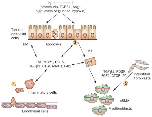
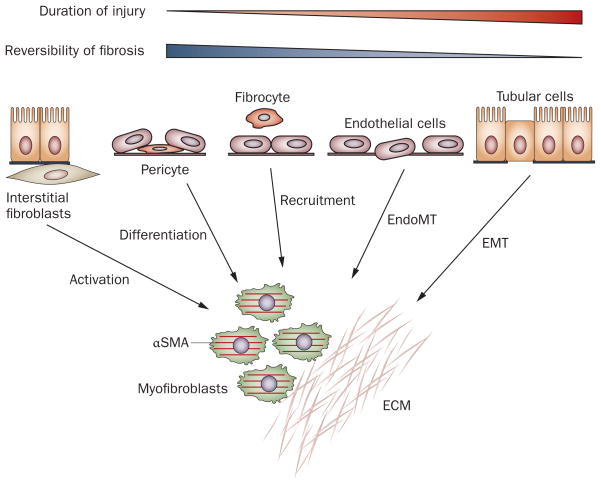
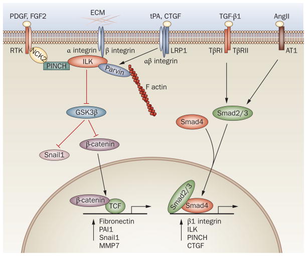
Renal fibrosis, particularly tubulointerstitial fibrosis, is the common final outcome of almost all progressive chronic kidney diseases. Renal fibrosis is also a reliable predictor of prognosis and a major determinant of renal insufficiency. Irrespective of the initial causes, renal fibrogenesis is a dynamic and converging process that consists of four overlapping phases: priming, activation, execution and progression. Nonresolving inflammation after a sustained injury sets up the fibrogenic stage (priming) and triggers the activation and expansion of matrix-producing cells from multiple sources through diverse mechanisms, including activation of interstitial fibroblasts and pericytes, phenotypic conversion of tubular epithelial and endothelial cells and recruitment of circulating fibrocytes. Upon activation, matrix-producing cells assemble a multicomponent, integrin-associated protein complex that integrates input from various fibrogenic signals and orchestrates the production of matrix components and their extracellular assembly. Multiple cellular and molecular events, such as tubular atrophy, microvascular rarefaction and tissue hypoxia, promote scar formation and ensure a vicious progression to end-stage kidney failure. This Review outlines our current understanding of the cellular and molecular mechanisms of renal fibrosis, which could offer novel insights into the development of new therapeutic strategies.


Conflict of interest statement

Competing interests

The author declares no competing interests.

Figures

Figure 1
Figure 1
Major events in renal interstitial fibrogenesis. (1) Peritubular infiltration of inflammatory cells, particularly T cells and macrophages, is an early event that sets up a fibrogenic stage. (2) Myofibroblast activation and expansion from various sources. The majority of the matrix-producing myofibroblasts are probably generated from local activation of interstitial fibroblasts. (3) Tubular cell apoptosis and EMT, leading to tubular atrophy. Abbreviations: αSMA, α smooth muscle actin; AngII, angiotensin II; CCL5, chemokine (C–C motif) ligand 5; CTGF, connective tissue growth factor; EMT, epithelial–mesenchymal transition; FGF2, basic fibroblast growth factor; MCP1, monocyte chemotactic protein 1; MMPs, matrix metalloproteinases; PAI1, plasminogen activator inhibitor 1; PDGF, platelet-derived growth factor; TBM, tubular basement membrane; TGF-β1, transforming growth factor β1; TNF, tumor necrosis factor; tPA, tissue-type plasminogen activator.
Figure 2
Figure 2
Multiple origins of myofibroblasts have been proposed in renal fibrosis. Myofibroblasts can be derived from at least five different sources through various mechanisms: phenotypic activation from interstitial fibroblasts; differentiation from vascular pericytes; recruitment from circulating fibrocytes; capillary EndoMT; and tubular EMT. The relative contribution of each source to the myofibroblast pool in renal fibrosis is controversial. Conceivably, local activation of resident fibroblasts remains the major route for the generation of myofibroblasts in diseased kidneys, at least in the early stage. By contrast, EMT could be a late event, and contribute to the irreversible progression of fibrosis. Abbreviations: αSMA, α smooth muscle actin; ECM, extracellular matrix; EMT, epithelial–mesenchymal transition; EndoMT, endothelial–mesenchymal transition.
Figure 3
Figure 3
A multicomponent, integrin-associated protein complex constitutes the molecular machinery that integrates various fibrogenic signals and orchestrates matrix production and assembly. Integrins and ILK and their associated proteins constitute the core components of this machinery. TGF-β1 regulates the expression of major components of this complex such as β1 integrin, ILK and PINCH via Smad signaling. AngII activates Smad3 signaling through pathways that are either dependent on or independent of TGF-β1, which leads to upregulation of the components of this complex. PDGF and FGF2 activate their respective receptor tyrosine kinases and influence this complex via cytoplasmic protein NCK2. CTGF and tPA bind to LRP1 and recruit β1 integrin, and then activate this machinery. This complex also activates β-catenin and Snail1 through inhibition of GSK3β, and thereby directly controls the expression of various fibrosis-related genes. The red lines indicate inhibition, the black lines indicate promotion. Abbreviations: AngII, angiotensin II; AT1, angiotensin II type I receptor; CTGF, connective tissue growth factor; ECM, extracellular matrix; FGF2, basic fibroblast growth factor; GSK3β, glycogen synthase kinase 3β; ILK, integrin-linked kinase; LRP1, LDL receptor-related protein 1; MMP7, matrix metalloproteinase 7; NCK2, non-catalytic region of tyrosine kinase adaptor protein 2; PAI1, plasminogen activator inhibitor 1; PDGF, platelet-derived growth factor; PINCH, particularly interesting new cysteine-histidine rich protein; RTK, receptor tyrosine kinase; TCF, T cell factor; TGF-β1, transforming growth factor β1; TβR1, type I TGF-β receptor; TβRII, type II TGF-β receptor; tPA, tissue-type plasminogen activator.

References

    1. Coresh J, et al. Prevalence of chronic kidney disease in the United States. JAMA. 2007;298:2038–2047. - PubMed
    1. United States Renal Data System. Annual Data Report 2009. 2010 [online], http://www.usrds.org/adr.htm.
    1. Sharma SK, et al. Burden of CKD, proteinuria, and cardiovascular risk among Chinese, Mongolian, and Nepalese participants in the International Society of Nephrology screening programs. Am J Kidney Dis. 2010;56:915–927. - PubMed
    1. Zhang L, et al. Prevalence and factors associated with CKD: a population study from Beijing. Am J Kidney Dis. 2008;51:373–384. - PubMed
    1. Liu Y. Renal fibrosis: new insights into the pathogenesis and therapeutics. Kidney Int. 2006;69:213–217. - PubMed

Publication types

Substances