. 2012 Feb;226(3):451-62.
doi: 10.1002/path.2986. Epub 2011 Oct 18.

The retinoid signalling molecule, TRIM16, is repressed during squamous cell carcinoma skin carcinogenesis in vivo and reduces skin cancer cell migration in vitro

Affiliations
Free PMC article

The retinoid signalling molecule, TRIM16, is repressed during squamous cell carcinoma skin carcinogenesis in vivo and reduces skin cancer cell migration in vitro

Belamy B Cheung et al. J Pathol. 2012 Feb.
Free PMC article

Abstract

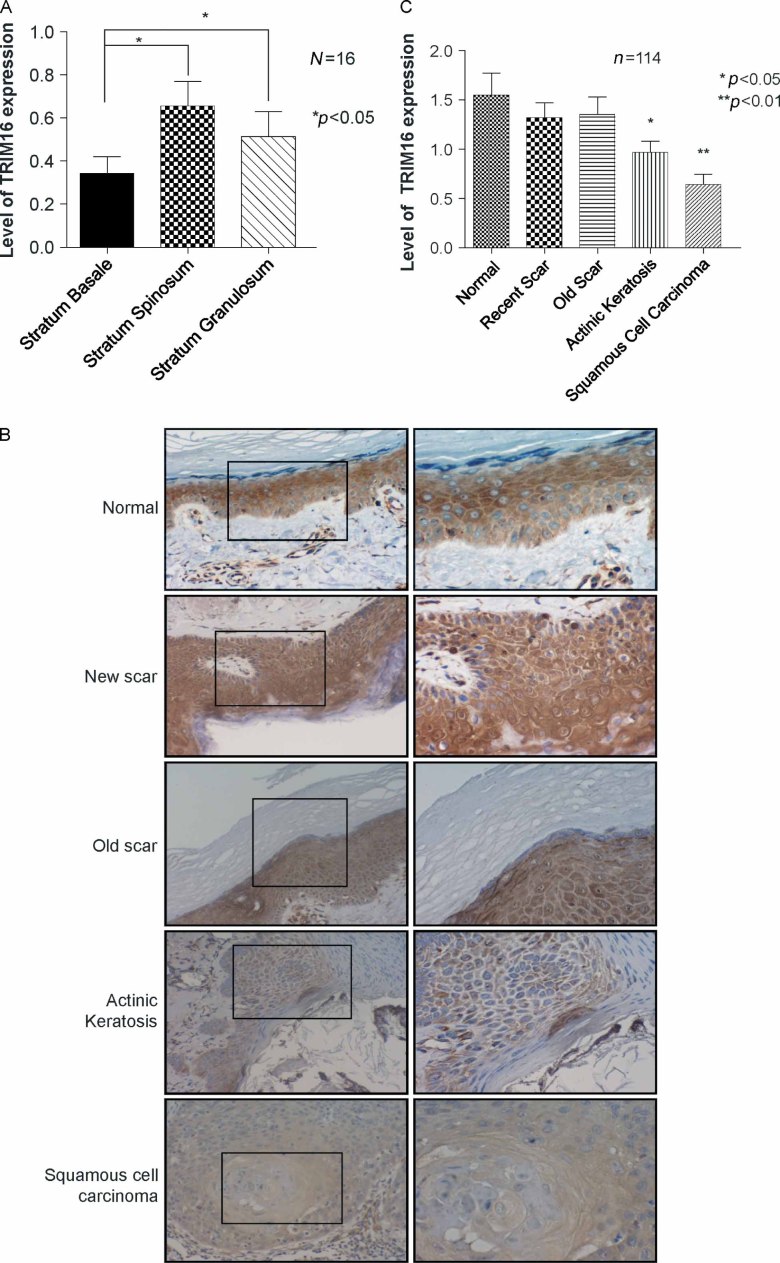
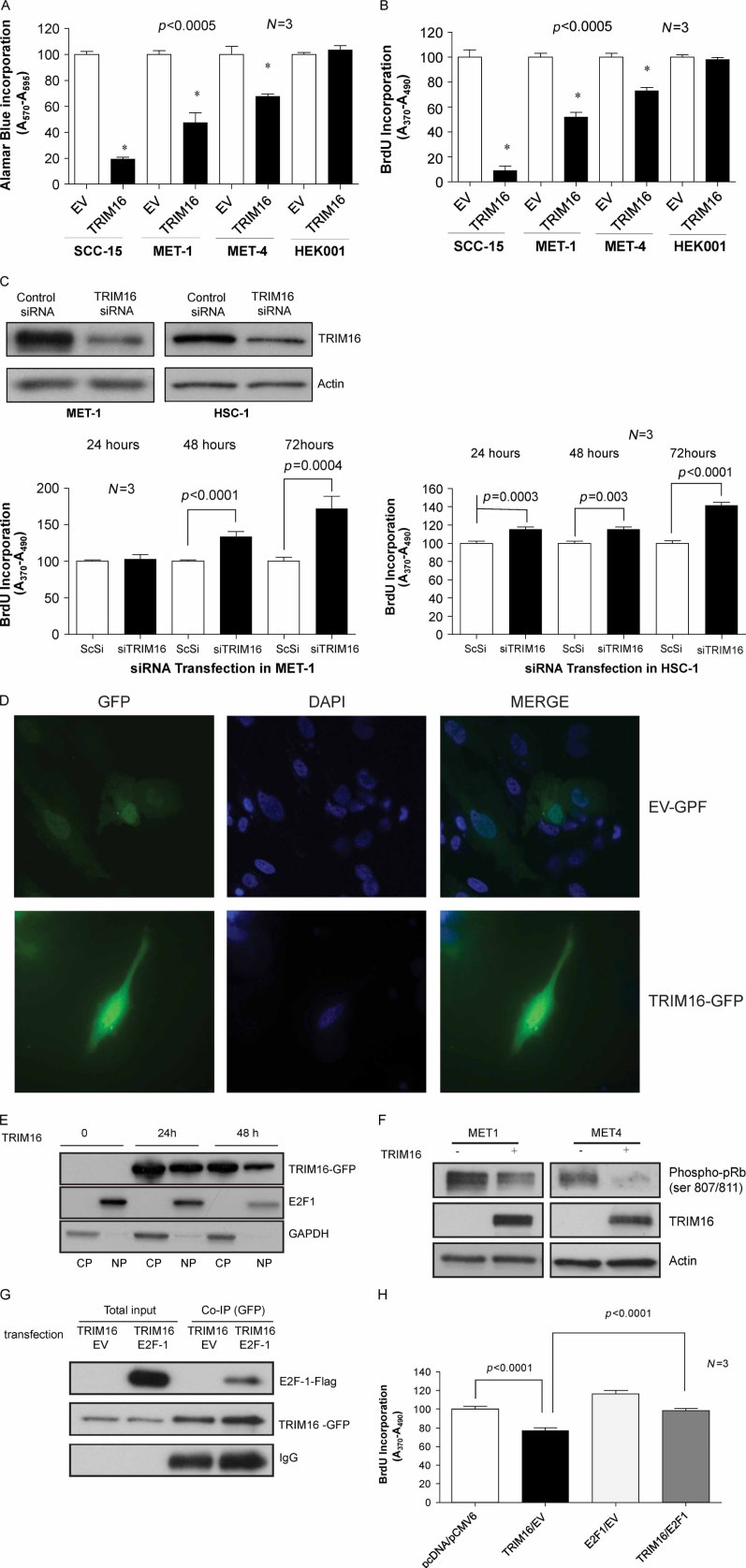
Retinoid therapy is used for chemo-prevention in immuno-suppressed patients at high risk of developing skin cancer. The retinoid signalling molecule, tripartite motif protein 16 (TRIM16), is a regulator of keratinocyte differentiation and a tumour suppressor in retinoid-sensitive neuroblastoma. We sought to determine the role of TRIM16 in skin squamous cell carcinoma (SCC) pathogenesis. We have shown that TRIM16 expression was markedly reduced during the histological progression from normal skin to actinic keratosis and SCC. SCC cell lines exhibited lower cytoplasmic and nuclear TRIM16 expression compared with primary human keratinocyte (PHK) cells due to reduced TRIM16 protein stability. Overexpressed TRIM16 translocated to the nucleus, inducing growth arrest and cell differentiation. In SCC cells, TRIM16 bound to and down regulated nuclear E2F1, this is required for cell replication. Retinoid treatment increased nuclear TRIM16 expression in retinoid-sensitive PHK cells, but not in retinoid-resistant SCC cells. Overexpression of TRIM16 reduced SCC cell migration, which required the C-terminal RET finger protein (RFP)-like domain of TRIM16. The mesenchymal intermediate filament protein, vimentin, was directly bound and down-regulated by TRIM16 and was required for TRIM16-reduced cell migration. Taken together, our data suggest that loss of TRIM16 expression plays an important role in the development of cutaneous SCC and is a determinant of retinoid sensitivity.


Figures

Figure 1
Figure 1
Reduced expression of TRIM16 in human skin SCC compared with normal skin. (A) The distribution of TRIM16 expression in normal epidermis: the data were collected from 16 normal patient skin samples; four fields were selected from each sample. (B) Representative sections showing immunohistochemical staining for TRIM16 expression (brown staining) in five categories of primary human skin tissues, using an anti-TRIM16 antibody (scale bar = 100 µm). Original magnification: left panels, 200×; right panels, 400×. (C) Analysis of the staining intensity of 114 patients showed that TRIM16 expression is markedly reduced during the progression from normal skin to SCC. Statistical analysis was performed using one-way analysis of variance
Figure 2
Figure 2
TRIM16 protein is regulated by the proteasome-dependent pathway and its expression level is reduced in SCC cells. (A) Immunoblotting analysis of the expression of cytoplasmic and nuclear proteins TRIM16 in PHKs, MET-1, MET-4, and SCC-15 cells. Anti-histone H3 was used as a control for nuclear protein expression, and the anti-GAPDH antibody as a cytoplasmic protein control. (B, C) MET-1, MET-4, and HEK001 cells were treated with cycloheximide at a final concentration of 100 µg/ml over 8 h. At the specified time points, the cells were harvested and the protein was extracted for analysis by western blots. The western blots were probed with anti-cyclin E2 and anti-actin antibodies as controls. (D) Cells were treated with 30 µm MG-132 for 5 h. Whole cell lysates were prepared and western blots were probed with anti-TRIM16, cyclin E2, and actin antibodies
Figure 3
Figure 3
Retinoid treatment increases the nuclear level of TRIM16 in PHKs, but not in SCC cells. (A, C) Immunoblotting analysis of cytoplasmic and nuclear TRIM16 proteins in PHKs and MET-1 cells treated with 1 µm 13-cis-RA or 10 µm 13-cis-RA for 24 and 48 h, respectively. Anti-histone H3 was used as a control for nuclear protein, and anti-GAPDH antibody as a cytoplasmic protein control. (B, D) PHKs and MET-1 cells were treated with the indicated concentrations of 13-cis-RA for 48 h, followed by incubation with BrdU for the last 6 h. BrdU incorporation was measured as OD units of absorbance
Figure 4
Figure 4
Increased TRIM16 expression induces differentiation in SCC cells. (A) Differentiated HaCaT cells were maintained in the presence of Ca2+ (+), and dedifferentiated cells in the absence of Ca2+ (−). Whole cell lysates of the HaCaT cells were subjected to the immunoblotting for involucrin, TRIM16, or actin. (B) Immunoblots of total cellular proteins from confluent HSC-1 cells transiently overexpressing TRIM16 plasmid DNA and control cells transfected with empty vector using cytokeratin 1/10, involucrin, myc-tag, and actin antibodies
Figure 5
Figure 5
Enforced overexpression caused TRIM16 translocation to the nucleus, reduced cell growth, and decreased nuclear E2F1 and pRb phosphorylation. (A, B) Cell viability and proliferation were measured at 72 h by Alamar Blue assay or BrdU incorporation, respectively. Cells were transiently transfected with empty vector (EV) or with TRIM16 plasmid DNA. (C) Top panel: western blotting analysis by TRIM16-specific antibody at 72 h of MET-1 and HSC-1 cells with control siRNA or TRIM16 siRNA transfection. Bottom panel: cell proliferation was measured up to 72 h by BrdU incorporation assay in MET-1 and HSC-1 cells transfected with control siRNA (ScSi) or TRIM16-specific siRNA (siTRIM16). (D) Immunofluorescence microscopy of nuclear TRIM16 staining in MET-1cells after empty vector and GFP-TRIM16 transfection for 24 h. TRIM16 protein was detected with fluorescein (green), and nuclei were identified with DAPI (blue). (E) Immunoblotting analysis of the expression of TRIM16 and E2F1 in MET-1 cells. Cytoplasmic (CP) and nuclear (NP) proteins of cells transfected with TRIM16 plasmid DNA for 24 and 48 h were analysed by immunoblotting using anti-TRIM16 and anti-E2F1 antibodies. (F) Phospho-pRb (ser807/811) protein was analysed by western blot with samples from MET-1 and MET-4 cells, transiently transfected with TRIM16 cDNA plasmid or empty vector. Actin was used as a loading control. GFP-tag antibody was probed for confirmation of TRIM16 plasmid transfection. (G) Lysates of MET-1 cells transfected with the indicated plasmids (lanes 1 and 3: GFP-TRIM16 and pCMV-6 empty vectors; lanes 2 and 4: GFP-TRIM16 and pCMV-6-E2F1) were immunoprecipitated by anti-GFP antibody and analysed by immunoblotting using anti-Flag and anti-GFP antibodies. (H) MET-1 cells were transfected with different plasmid DNAs for 72 h, followed by incubation with BrdU for the last 6 h
Figure 6
Figure 6
TRIM16 binds to and modulates vimentin protein expression in SCC cells. (A) Interaction of TRIM16 with vimentin. Lysates of MET-1 cells transfected with either TRIM16-GFP or vimentin-Flag plasmid DNA and immunoprecipitated with GFP antibody were then analysed by immunoblotting using anti-GFP and anti-Flag tag antibodies. (B) TRIM16 overexpression down-regulates exogenous vimentin. Lysates of TRIM16 or empty vector transfected MET-1 cells were analysed by anti-GFP and anti-vimentin antibodies. Anti-actin antibody served as a loading control
Figure 7
Figure 7
Overexpression of TRIM16 reduces cell motility and migration through its RFP-like domain. (A) Representative phase contrast micrographs of closure of scratch-wounded confluent cultures of empty vector or TRIM16 plasmid DNA transfected MET-1 and MET-4 cells after wounding and 8 h post-wounding. (B) Relative closure of MET-1 and MET-4 wounds: the average distance moved by the empty vector control and the average (and standard error) movement of TRIM16 plasmid DNA transfected cells in three independent wounds are shown relative to percentage of original wound. (C) Representative phase contrast micrographs of scratch-wounded confluent cultures of empty vector or TRIM16 full-length, M1, M2, M3, and M4 plasmid DNA transfected MET-1 cells after wounding and 24 h post-wounding. (D) Relative closure of MET-1 wounds: the average distance moved by the empty vector control and the average (and standard error) movement of TRIM16 and M1, M2, M3 and M4 transfected cells in three independent wounds are shown relative to percentage of original wound. (E) Schematic representations of TRIM16 full-length, mutant 1 (M1), mutant 2 (M2), mutant 3 (M3), and mutant 4 (M4) constructs used in this study. (F) Expression of TRIM16 full-length and deletion mutants in MET-1 cells: the lysates of MET-1 cells transiently transfected with empty vector (EV, lane 1), TRIM16 full-length (lane 2, 95 kD), mutant 1 (lane 3, 70 kD), mutant 2 (lane 4, 55 kD), mutant 3 (lane 5, 65 kD), and mutant 4 (lane 6, 45 kD) were probed with the anti-GFP tag antibody in an immunoblot analysis. (G) Invasion assay of MET-1 cells through collagen-coated cell culture inserts. The cells were transiently transfected with empty vector, TRIM16 full-length, mutant 3, and mutant 4 for 24 h. The percentage of the migrated cells divided by the total number of cells in the wells is shown. (H) Invasion assay of MET-1 cells through collagen-coated cell culture inserts. The cells were transiently transfected with empty vector, TRIM16 full-length, TRIM16 and vimentin, or vimentin alone for 24 h. The percentage of migrated cells divided by the total number of cells in the wells from three independent experiments is shown

Similar articles

Cited by

References

    1. Lansbury L, Leonardi-Bee J, Perkins W, et al. Interventions for non-metastatic squamous cell carcinoma of the skin. Cochrane Database Syst Rev. 2010;4:CD007869. - PMC - PubMed
    1. Armstrong BK, Kricker A. The epidemiology of UV induced skin cancer. J Photochem Photobiol B. 2001;63:8–18. - PubMed
    1. Halliday GM, Norval M, Byrne SN, et al. The effects of sunlight on the skin. Drug Discov Today: Disease Mechanisms. 2008;5:e201–e209.
    1. Lotan R, Xu XC, Lippman SM, et al. Suppression of retinoic acid receptor-beta in premalignant oral lesions and its up-regulation by isotretinoin. N Engl J Med. 1995;332:1405–1410. - PubMed
    1. Cheung B, Hocker JE, Smith SA, et al. Retinoic acid receptors beta and gamma distinguish retinoid signals for growth inhibition and neuritogenesis in human neuroblastoma cells. Biochem Biophys Res Commun. 1996;229:349–354. - PubMed

Publication types

MeSH terms