. 2012 Feb;40(3):1356-65.
doi: 10.1093/nar/gkr843. Epub 2011 Oct 18.

Molecular basis of differential target regulation by miR-96 and miR-182: the Glypican-3 as a model

Affiliations

Molecular basis of differential target regulation by miR-96 and miR-182: the Glypican-3 as a model

Sandra Jalvy-Delvaille et al. Nucleic Acids Res. 2012 Feb.

Abstract

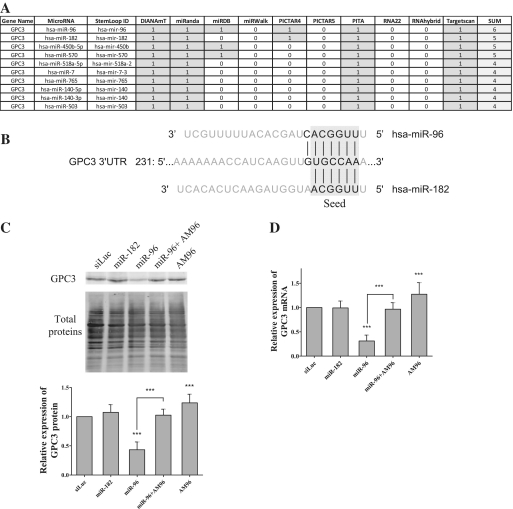
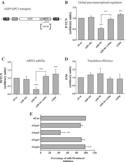
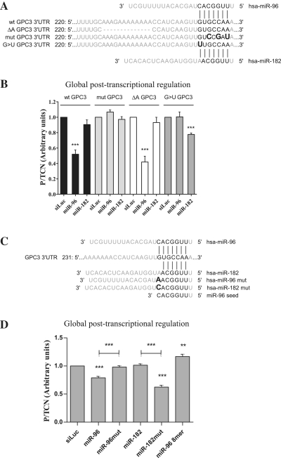
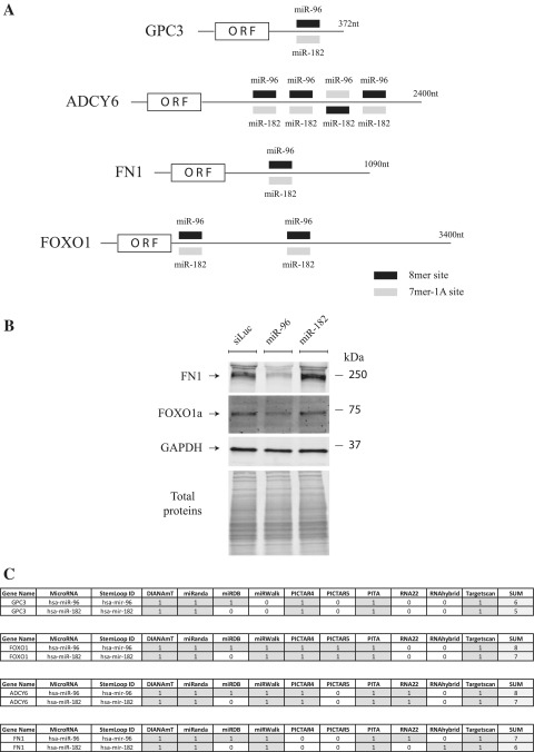
Besides the fact that miR-96 and miR-182 belong to the miR-182/183 cluster, their seed region (UUGGCA, nucleotides 2-7) is identical suggesting potential common properties in mRNA target recognition and cellular functions. Here, we used the mRNA encoding Glypican-3, a heparan-sulfate proteoglycan, as a model target as its short 3' untranslated region is predicted to contain one miR-96/182 site, and assessed whether it is post-transcriptionally regulated by these two microRNAs. We found that miR-96 downregulated GPC3 expression by targeting its mRNA 3'-untranslated region and interacting with the predicted site. This downregulatory effect was due to an increased mRNA degradation and depended on Argonaute-2. Despite its seed similarity with miR-96, miR-182 was unable to regulate GPC3. This differential regulation was confirmed on two other targets, FOXO1 and FN1. By site-directed mutagenesis, we demonstrated that the miRNA nucleotide 8, immediately downstream the UUGGCA seed, plays a critical role in target recognition by miR-96 and miR-182. Our data suggest that because of a base difference at miRNA position 8, these two microRNAs control a completely different set of genes and therefore are functionally independent.


Figures

Figure 1.
Figure 1.
GPC3 is a target of miR-96, but not of miR-182. (A) The 10 highest-ranking miRNA:GPC3-3′-UTR pairings as predicted by miRWalk using the indicated algorithms (1/grey: pairing predicted; 0/empty: pairing not predicted). (B) Schematic representation of the pairing of GPC3 3′-UTR with miR-96 or miR-182 as predicted by TargetScan. In this figure and the following ones, base pairings between indicated mRNAs and miRNAs are represented by vertical lines. (C and D): HuH7 cells were transfected with small RNAs as indicated. Three days later, the amounts of GPC3 protein (C) and mRNA (D) were quantified (ANOVA: P < 0.0001; n = 6). In Panel C, a representative western blot experiment is shown on top and a bar graph recapitulating means with standard deviation (SD) of six experiments is shown on bottom. In this figure and the following ones, bars represent means, error bars indicate SD and the ANOVA test was followed by a Bonferroni's multiple comparison post-test. ***P < 0.001.
Figure 2.
Figure 2.
Molecular basis of the post-transcriptional regulation mediated by miR-96 on GPC3. (A) Schematic representation of the eGFP-GPC3 transgene with the 3′-UTR being targeted by miR-96. (B–D) HuH7 cells were transduced once with lentiviral particles expressing the transgene. After one week, the TCN was calculated using genomic DNA extracted from the eGFP-GPC3-expressing HuH7 cell population. Then cells were transfected with the indicated small RNAs. Three days later, the eGFP protein expression and mRNA amount were measured and data were analyzed following the FunREG experimental pipeline (9). (B) Global post-transcriptional regulation (ANOVA: P < 0.0001; n = 6). (C) mRNA stability (ANOVA: P < 0.0001; n = 6). (D) Translation efficiency (ANOVA: P = NS; n = 6). Panel (E): EGFP-GPC3-expressing HuH7 cells were first transfected with the indicated siRNA. Twenty-four hours later, cells were transfected with miR-96. Forty-eight hours later, eGFP protein expression was analyzed by FACS and data normalized to that of siLuc (ANOVA: P < 0.0001; n = 3). ***P < 0.001.
Figure 3.
Figure 3.
Base requirements for miR-96:GPC3 3′-UTR recognition and miR-96 function. (A) Schematic representation of miR-96 or miR-182 pairing with the GPC3 3′-UTR sequence in its wild-type, deleted or mutated versions. (B) HuH7 cells expressing the indicated transgene (shown above the bars; TCN value measured) were transfected with the indicated small RNAs. Three days later, eGFP protein expression was analyzed by FACS as described in Figure 2B (ANOVA: P < 0.0001; n = 3). (C) Schematic representation of the wild type GPC3 3′-UTR sequence and the various synthetic double-strand small RNAs used (only the guide strand is shown). (D) eGFP-GPC3-expressing HuH7 cells were transfected with the indicated small RNAs. Three days later, eGFP protein expression was analyzed as described in panel B (ANOVA: P < 0.0001; n = 5). **P < 0.01; ***P < 0.001.
Figure 4.
Figure 4.
Target preference of miR-96 and miR-182 depends on presence of a 8-mer site. (A) Schematic representation of the indicated mRNAs with their miR-96/182 sites. The type of site as defined by Targetscan is as indicated (see the legend at the bottom-right). (B) HuH7 cells were transfected with small RNAs as indicated. Three days later, the amounts of FN1, FOXO1 and GAPDH proteins were measured. Representative of three independent experiments. (C) miRNA:target 3′-UTR pairings as predicted by miRWalk using the indicated programs.

Similar articles

Cited by

References

    1. Barreau C, Paillard L, Osborne HB. AU-rich elements and associated factors: are there unifying principles? Nucleic Acids Res. 2005;33:7138–7150. - PMC - PubMed
    1. Garneau NL, Wilusz J, Wilusz CJ. The highways and byways of mRNA decay. Nat. Rev. Mol. Cell Biol. 2007;8:113–126. - PubMed
    1. Grosset C, Chen CY, Xu N, Sonenberg N, Jacquemin-Sablon H, Shyu AB. A mechanism for translationally coupled mRNA turnover: interaction between the poly(A) tail and a c-fos RNA coding determinant via a protein complex. Cell. 2000;103:29–40. - PubMed
    1. Grosset C, Boniface R, Duchez P, Solanilla A, Cosson B, Ripoche J. In vivo studies of translational repression mediated by the granulocyte-macrophage colony-stimulating factor AU-rich element. J. Biol. Chem. 2004;279:13354–13362. - PubMed
    1. Bartel DP. MicroRNAs: target recognition and regulatory functions. Cell. 2009;136:215–233. - PMC - PubMed

Publication types