. 2012 Feb;40(3):1381-94.
doi: 10.1093/nar/gkr890. Epub 2011 Oct 18.

Analysis of the interaction with the hepatitis C virus mRNA reveals an alternative mode of RNA recognition by the human La protein

Affiliations

Analysis of the interaction with the hepatitis C virus mRNA reveals an alternative mode of RNA recognition by the human La protein

Luigi Martino et al. Nucleic Acids Res. 2012 Feb.

Abstract

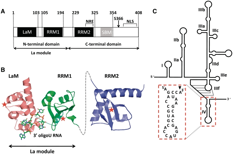
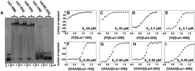
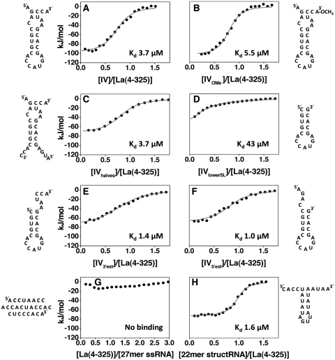
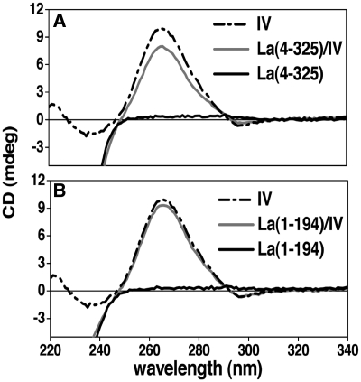
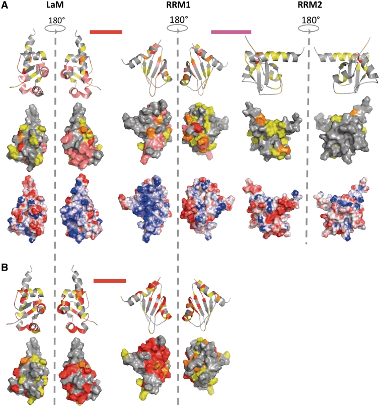
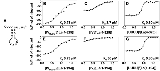
Human La protein is an essential factor in the biology of both coding and non-coding RNAs. In the nucleus, La binds primarily to 3' oligoU containing RNAs, while in the cytoplasm La interacts with an array of different mRNAs lacking a 3' UUU(OH) trailer. An example of the latter is the binding of La to the IRES domain IV of the hepatitis C virus (HCV) RNA, which is associated with viral translation stimulation. By systematic biophysical investigations, we have found that La binds to domain IV using an RNA recognition that is quite distinct from its mode of binding to RNAs with a 3' UUU(OH) trailer: although the La motif and first RNA recognition motif (RRM1) are sufficient for high-affinity binding to 3' oligoU, recognition of HCV domain IV requires the La motif and RRM1 to work in concert with the atypical RRM2 which has not previously been shown to have a significant role in RNA binding. This new mode of binding does not appear sequence specific, but recognizes structural features of the RNA, in particular a double-stranded stem flanked by single-stranded extensions. These findings pave the way for a better understanding of the role of La in viral translation initiation.


Figures

Figure 1.
Figure 1.
Details of the human La protein and the HCV IRES domain. (A) Domain organization of human La showing the La motif (LaM) and RRM1 (forming the La module), the RRM2, the nuclear retention element (NRE), the Short Basic Motif (SBM) and nuclear localization signal (NLS). (B) Structured domains of human La, depicting the crystal structure of the La module in complex with a 3′ oligoU ssRNA (PDB ID 2VOP) and the solution structure of the isolated RRM2 (PDB ID 1OWX). A dotted grey line denotes the linker between the RRM1 and RRM2. Red stars indicate the canonical RNA-binding surfaces on the three domains. (C) Schematic representation of the secondary structure elements and domains of the HCV IRES. The domain fragment used in biophysical experiments (domain IV) is highlighted. The black arrowhead indicates the base that was mutated from U to A in our experiments.
Figure 2.
Figure 2.
Interaction of the human La protein with domain IV of the HCV IRES. (A) Representative EMSA experiment of domain IV RNA (20 nM) mixed with two different concentrations (0.5 and 2.0 µM) of the following proteins: full-length La(1–408), La(1–354), La(4–325), La(1–194) and La(105–325). (B, C, D, E) ITC experiments of the interaction of domain IV RNA with La(1–194), La(1–229), La(4–325) and La(1–408) respectively. The normalized heat of interaction for the titrations was obtained by integrating the raw data and subtracting the heat of ligand dilution into the buffer alone. The grey line represents the best fit obtained by a non-linear least-squares procedure based on an independent binding sites model. Dissociation constants (Kd) are shown for each interaction. The binding profile observed for the full-length protein is unchanged in the mutant La(4–325), but a dramatic decrease in binding affinity is observed for mutants lacking RRM2. (F, G, H, I) ITC experiments of the interaction of UUUUOH RNA with La(1–194), La(1–229), La(4–325) and La(1–408). All the proteins show similar affinities and thermodynamic profiles for the interaction with this RNA, confirming that La(1–194) is sufficient for binding to short 3′ U-rich RNA sequences.
Figure 3.
Figure 3.
Interaction of La(4–325) with RNA targets. (A–H) ITC experiments showing the thermal effect of mixing La(4–325) with different RNA targets as follows: (A) IV; (B) IVOMe; (C) IVhalves; (D) IVlowerSL; (E) IV3′ext; (F) IV5′ext; (G) 27-mer ssRNA; (H) 22-mer structRNA. The grey lines represent the best fit derived by a non-linear least-squares procedure based on an independent binding sites model. The expected secondary structures of the RNA molecules obtained with the software mfold, which matched well with CD thermal unfolding data (Supplementary Figure S1), are shown beside each graph. Taken together, these data show that La(4–325) recognizes a double-stranded stem flanked by extensions at the 5′ or 3′, independently of nucleotide sequence.
Figure 4.
Figure 4.
CD analysis of domain IV conformation upon interaction with La. (A) Near-UV CD spectra of domain IV RNA alone (dotted line) and in complex with the La(4–325) (grey line) show a modest reduction in the intensity of the CD signal at 263 nm following protein binding. Since the signal from the RNA dominates the CD spectrum at wavelengths >250 nm (near UV), with the signal of the protein being negligible in this range (black line), any spectral difference here could therefore be attributed solely to changes in the polynucleotide conformation. (B) Near-UV CD spectra of domain IV RNA alone (dotted line) and in complex with the La(1–194) (grey line). In this case a much smaller change in the RNA CD signal was detected.
Figure 5.
Figure 5.
NMR analysis of La-domain IV interaction. (A) Structure mapping of the chemical shift variations obtained in La(4–325) on domain IV binding for the LaM, RRM1, RRM2 and inter-domain linkers and comparison with the electrostatic surface potential. The LaM, RRM1 and RRM2 are shown as structures of individual domains (PDB ID: 1S7A, 1S79 and 1OWX respectively), whereas the inter-domain linkers are shown as bars between domains. The positions of the residues that show substantial shifts are indicated on the protein secondary structures (top) and surfaces (middle). The chemical shift perturbations (ΔδAV) are represented with the following colour code: yellow (0.035 ≤ ΔδAV ≤ 0.06), orange (0.06 < ΔδAV ≤ 0.1), red (ΔδAV > 0.1), pink (disappearing signals) and magenta (not quantified) (Supplementary Figure S2C). (B) Structure mapping of the chemical shift variations obtained in La(1–194) on 3′ oligoU binding for the LaM, RRM1 and inter-domain linker.
Figure 6.
Figure 6.
Comparative analysis of the interaction between La and IVUUUU RNA. (A) Sequence and secondary structure for IVUUUU, which retains the lower stem–loop portion of domain IV while the 5′ and 3′ extensions are mutated to oligoC and oligoU, respectively. (B and E) ITC experiments examining the interaction of IVUUUU with La(4–325) and La(1–194) respectively. The grey line represents the best fit obtained by a non-linear least-squares procedure based on an independent binding sites model. For comparison, the interactions of domain IV (C and F) and UUUUOH (D and G) with La(4–325) and La(1–194) respectively are reported. For the interactions of La(4–325) and La(1–194) with IVUUUU, the thermodynamic signature of binding, evaluated as the relative ΔH/–TΔS value, is identical in both cases and is closer to the UUUUOH associations (Table 1).

References

    1. Wolin SL, Cedervall T. The La protein. Annu. Rev. Biochem. 2002;71:375–403. - PubMed
    1. Bayfield MA, Yang R, Maraia RJ. Conserved and divergent features of the structure and function of La and La-related proteins (LARPs) Biochim. Biophys. Acta. 2010;1799:365–378. - PMC - PubMed
    1. Xue D, Rubinson DA, Pannone BK, Yoo CJ, Wolin SL. U snRNP assembly in yeast involves the La protein. EMBO J. 2000;19:1650–1660. - PMC - PubMed
    1. Kufel J, Allmang C, Chanfreau G, Petfalski E, Lafontaine DL, Tollervey D. Precursors to the U3 small nucleolar RNA lack small nucleolar RNP proteins but are stabilized by La binding. Mol. Cell. Biol. 2000;20:5415–5424. - PMC - PubMed
    1. Yoo CJ, Wolin SL. The yeast La protein is required for the 3′ endonucleolytic cleavage that matures tRNA precursors. Cell. 1997;89:393–402. - PubMed

Publication types