

Recurrence analysis in 66 cases with grade I and grade II chondrosarcomas in the extremities
- PMID: 22009830
- PMCID: PMC6583385
- DOI: 10.1111/j.1757-7861.2009.00020.x
Recurrence analysis in 66 cases with grade I and grade II chondrosarcomas in the extremities
Abstract
Objective: We analyzed chondrosarcomas in long bones to assess whether Grade I and II chondrosarcomas should both be grouped as low grade malignant tumors (musculoskeletal Tumor Society Stage I or Stage II), and to explore rational surgical treatment for Grade I and II chondrosarcomas.
Methods: We retrospectively reviewed 66 patients from January 1996 to December 2007 with Grade I and II chondrosarcoma of the extremities without metastases at the Department of Orthopaedics of Shanghai Sixth People's Hospital. Thirty-eight patients had undergone intralesional or marginal resection, and 28 patients had undergone wide marginal or radical excision based on imaging findings. The mean age of the patients was 43 years (range, 5-85) and the minimum follow-up was 31 months (mean, 48; range, 5-141). We analyzed grade diagnosis, therapeutic options, and local recurrence rate of the two grades of chondrosarcoma.
Results: Of all patients, 22 experienced local recurrence, making the local recurrence rate 33.3%. A statistically significant difference in outcome between patients with Grade I and Grade II tumors undergoing intralesional resection was identified. No significant difference according to surgical method was found between the two groups in total.
Conclusion: Grade II chondrosarcomas should be grouped as high stage malignant tumors (Stage II) and grade I chondrosarcomas assigned to the low stage malignant tumor group (Stage I). Our experience suggests the surgical method should be related to radiographic margin status and oncologic classification. Wide resection should be considered for Grade II, while intralesional resection is suitable for Grade I.
© 2009 Tianjin Hospital and Blackwell Publishing Asia Pty Ltd.
Figures

 ) indicates the site after curettage and allogeneic bone graft.) indicates the site after curettage and allogeneic bone graft.
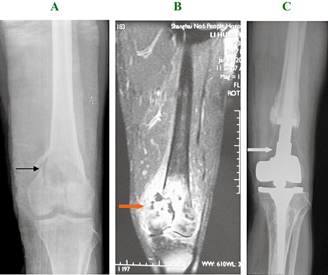
 ) indicates the lesion before surgery. (C) A 25‐month postoperative X‐ray image showing the appearance after treatment by wide marginal excision and artificial prosthetic replacement. There is no evidence of local recurrence. (
 ) indicates the prosthetic replacement.


References
-
- Gelderblom H, Hogendoorn PC, Dijkstra SD, et al. The clinical approach towards chondrosarcoma. Oncologist, 2008, 13: 320–329. - PubMed
-
- Evans HL, Ayala AG, Romsdahl MM. Prognostic factors in chondrosarcoma of bone: A clinicopathologic analysis with emphasis on histologic grading. Cancer, 1977, 40: 818–831. - PubMed
-
- Reith JD, Horodyski MB, Scarborough MT. Grade 2 chondrosarcoma: stage I or stage II tumor? Clin Orthop Relat Res, 2003, 415: 45–51. - PubMed
-
- Skeletal Lesions Interobserver Correlation among Expert Diagnosticians (SLICED) Study Group . Reliability of histopathologic and radiologic grading of cartilaginous neoplasms in long bones. J Bone Joint Surg Am, 2007, 89: 2113–2123. - PubMed
-
- Sanerkin NG. The diagnosis and grading of chondrosarcoma of bone: a combined cytologic and histologic approach. Cancer, 1980, 45: 582–594. - PubMed
Publication types
MeSH terms
LinkOut - more resources
Full Text Sources
Medical
