

Clinical, radiographic, and histological findings of florid cemento-osseous dysplasia: a case report
- PMID: 22010072
- PMCID: PMC3189539
- DOI: 10.5624/isd.2011.41.3.139
Clinical, radiographic, and histological findings of florid cemento-osseous dysplasia: a case report
Abstract
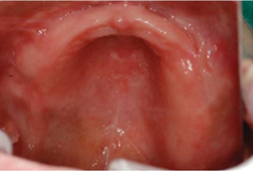
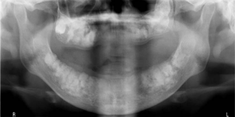
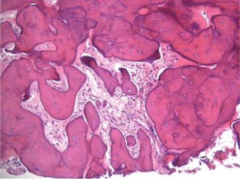
Cemento-osseous dysplasias are a group of disorders known to originate from periodontal ligament tissue and involve, essentially, the same pathological process. They are usually classified into three main groups: periapical, florid, and focal cemental dysplasias depending on their extent and radiographic appearances. Radiographically, florid cementoosseous dysplasia (FCOD) appears as dense, lobulated masses, often symmetrically located in various regions of the jaws. The best management for the asymptomatic FCOD patient consists of regular recall examinations with prophylaxis. The management of the symptomatic patient is more difficult. A case of FCOD occurring in a 52-year-old edentulous Korean female is reported which is rare with regard to race and sex.
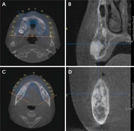
Keywords: Cementoma; Cone-Beam Computed Tomography; Florid Cemento-Osseous Dysplasia.
Figures
References
-
- Ong ST, Siar CH. Florid cemento-osseous dysplasia in a young Chinese man. Case report. Aust Dent J. 1997;42:404–408. - PubMed
-
- Miyake M, Nagahata S. Florid cemento-osseous dysplasia. Report of a case. Int J Oral Maxillofac Surg. 1999;28:56–57. - PubMed
-
- Waldron CA. Fibro-osseous lesions of the jaws. J Oral Maxillofac Surg. 1985;43:249–262. - PubMed
-
- Cannon JS, Keller EE, Dahlin DC. Gigantiform cementoma: report of two cases (mother and son) J Oral Surg. 1980;38:65–70. - PubMed
-
- Oikarinen K, Altonen M, Happonen RP. Gigantiform cementoma affecting a Caucasian family. Br J Oral Maxillofac Surg. 1991;29:194–197. - PubMed
