. 2012 Jan;340(1):176-84.
doi: 10.1124/jpet.111.187427. Epub 2011 Oct 18.

Impaired wound healing in hypoxic renal tubular cells: roles of hypoxia-inducible factor-1 and glycogen synthase kinase 3β/β-catenin signaling

Affiliations

Impaired wound healing in hypoxic renal tubular cells: roles of hypoxia-inducible factor-1 and glycogen synthase kinase 3β/β-catenin signaling

Jianping Peng et al. J Pharmacol Exp Ther. 2012 Jan.

Abstract

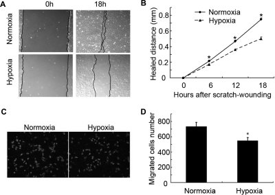
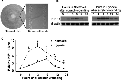
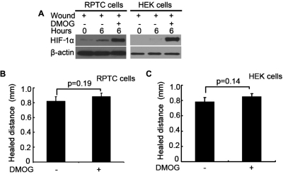
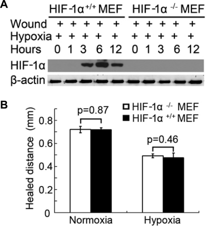
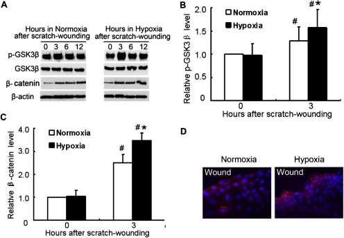
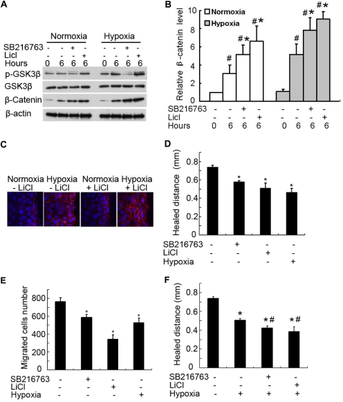
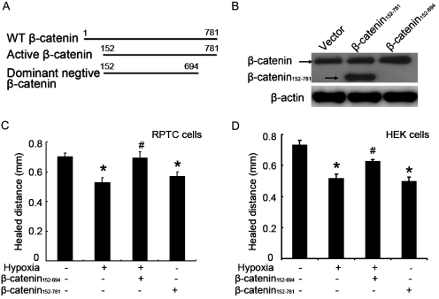
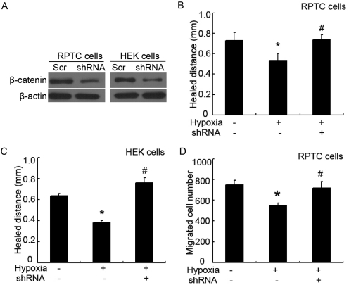
Wound and subsequent healing are frequently associated with hypoxia. Although hypoxia induces angiogenesis for tissue remodeling during wound healing, it may also affect the healing response of parenchymal cells. Whether and how wound healing is affected by hypoxia in kidney cells and tissues is currently unknown. Here, we used scratch-wound healing and transwell migration models to examine the effect of hypoxia in cultured renal proximal tubular cells (RPTC). Wound healing and migration were significantly slower in hypoxic (1% oxygen) RPTC than normoxic (21% oxygen) cells. Hypoxia-inducible factor-1α (HIF-1α) was induced during scratch-wound healing in normoxia, and the induction was more evident in hypoxia. Nevertheless, HIF-1α-null and wild-type cells healed similarly after scratch wounding. Moreover, activation of HIF-1α with dimethyloxalylglycine in normoxic cells did not suppress wound healing, negating a major role of HIF-1α in wound healing in this model. Scratch-wound healing was also associated with glycogen synthase kinase 3β (GSK3β)/β-catenin signaling, which was further enhanced by hypoxia. Pharmacological inhibition of GSK3β resulted in β-catenin expression, accompanied by the suppression of wound healing and transwell cell migration. Ectopic expression of β-catenin in normoxic cells could also suppress wound healing, mimicking the effect of hypoxia. Conversely, inhibition of β-catenin via dominant negative mutants or short hairpin RNA improved wound healing and transwell migration in hypoxic cells. The results suggest that GSK3β/β-catenin signaling may contribute to defective wound healing in hypoxic renal cells and tissues.


Figures

Fig. 1.
Fig. 1.
Hypoxia inhibits scratch-wound healing and transwell migration in RPTC. A and B, scratch-wound healing. RPTC were scratch-wounded with a sterile pipette tip and then incubated under hypoxia (1% O2) or normoxia (21% O2). A, representative wounds immediately after scratch wounding and after 18 h of healing were recorded with a phase-contrast microscope. B, the wound width was measured at 6, 12, and 18 h after scratching to determine the healed distance. C and D, transwell migration. A total of 3 × 10⁵ cells were added to each transwell insert, which was put in a 24-well plate with 600 μl of culture medium in normoxia or hypoxia for 6 h. C, representative PI staining of migratory cells was recorded with a fluorescence microscope. D, migratory cells attached on the undersurface were counted after PI staining. In B and D, data are expressed as mean ± S.D. (n = 6). *, p < 0.05 versus normoxia.
Fig. 2.
Fig. 2.
HIF-1α induction during wound healing in normoxia and hypoxia. A, multiple uniform wounds made with a wounding device. RPTC grown in 60-mm dishes were pressed with a multiple-wounding device to induce concentric circular cell bands of 130-μm width separated by wounds of 750-μm width. B, time-dependent HIF-1α induction during wound healing in normoxia and hypoxia. RPTC in 60-mm dishes were wounded by the multiple-wounding device and then incubated in normoxia or hypoxia to collect whole-cell lysates at different time points for immunoblot analysis of HIF-1α and β-actin. C, densitometric analysis of HIF-1α expression during scratch-wound healing. Immunoblots from three separate experiments were analyzed by densitometry, and the signal of HIF-1α was normalized with control (0 h) that was arbitrarily set as 1. Data are expressed as mean ± S.D. (n = 3). *, p < 0.05 versus normoxia.
Fig. 3.
Fig. 3.
DMOG induces HIF-1α without affecting wound healing. A, RPTC and HEK cells were wounded with the multiple-wounding device and incubated in full medium without (−) or with (+) 100 mM DMOG in normoxia for 6 h to collect whole-cell lysates for immunoblot analysis of HIF-1α. B and C, scratch-wound healing. RPTC and HEK cells were scratch-wounded and then incubated in normoxia without (−) or with (+) 100 mM DMOG for 18 h to measure the healed distance. Data are expressed as mean ± S.D. (n = 6).
Fig. 4.
Fig. 4.
Wound healing in HIF-1α-null cells. A, HIF-1α induction during wound healing in hypoxia in HIF-1α-null [HIF-1α(−/−)] and wild-type [(HIF-1α(+/+)] MEF cells. MEF cells were wounded by the multiple-wound device and incubated in hypoxia to collect whole-cell lysates at different time points for immunoblot analysis of HIF-1α and β-actin. B, scratch-wound healing. Wild-type and HIF-1α-null MEF cells were scratch-wounded and then incubated in hypoxia for 18 h to measure the healed distance. Data are expressed as mean ± S.D. (n = 6).
Fig. 5.
Fig. 5.
GSK3β and β-catenin expression during wound healing in RPTC. A, GSK3β phosphorylation and β-catenin induction during wound healing in RPTC under normoxia and hypoxia. RPTC were wounded by the multiple-wound device and incubated in normoxia or hypoxia to collect whole-cell lysates at different time points for immunoblot analysis of serine-15-phosphorylated and total GSK3β, β-catenin, and β-actin. B and C, densitometric analysis of p-GSK3β and β-catenin induction during 3 h of wound healing. The signals of p-GSK3β and β-catenin in the immunoblots from three separate experiments were analyzed by densitometry and normalized with control (0 h). Data are expressed as mean ± S.D. (n = 3). #, p < 0.05 versus control; *, p < 0.05 versus 3 h after scratch wounding in normoxia. D, immunofluorescence analysis of β-catenin expression. PRTC cells were subjected to 6 h of scratch-wound healing under normoxia or hypoxia. The cells were fixed for immunofluorescent staining of β-catenin (red) and nuclear staining with Hoechst33342 (blue).
Fig. 6.
Fig. 6.
GSK3β inhibitors suppress wound healing. A and B, β-catenin up-regulation by GSK3β inhibitors. RPTC were scratch-wounded and then incubated for 6 h in normoxia or hypoxia with or without 10 mM LiCl or 10 μM SB216763. A, whole-cell lysate was collected for immunoblot analysis of serine-9-phosphorylated GSK3β, total GSK3β, β-catenin, and β-actin. B, densitometric analysis of β-catenin expression. The signal of β-catenin in the immunoblots from three separate experiments was analyzed by densitometry and normalized with control (0 h). Data are expressed as mean ± S.D. (n = 3). #, p < 0.05 versus control; *, p < 0.05 versus 6 h of scratch-wound healing without inhibitors. C, another group of cells was fixed for immunofluorescent staining of β-catenin (red) and nuclear staining with Hoechst33342 (blue). D, suppression of wound healing in normoxia by GSK3β inhibitors. RPTC were scratch-wounded and incubated in normoxia with or without 10 mM LiCl and 10 μM SB216763 for 18 h to measure the healed distance. E, suppression of cell migration in normoxia by GSK3β inhibitors. A total of 3 × 10⁵ RPTC were seeded in a transwell insert, which was put in a 24-well plate containing 600 μl of medium with or without 10 mM LiCl and 10 μM SB216763 in normoxia for 6 h. The cells that migrated to the undersurface of the insert were stained with PI and counted. F, effect of GSK3β inhibitors on wound healing in hypoxia. RPTC were scratch-wounded and incubated in hypoxia with or without 10 mM LiCl and 10 μM SB216763 for 18 h to measure the healed distance. In D to F, data are expressed as mean ± S.D. (n = 6). *, p < 0.05 versus control; #, p < 0.05 versus hypoxia-only group.
Fig. 7.
Fig. 7.
Active β-catenin suppresses and dominant negative β-catenin enhances wound healing. RPTC and HEK cells were infected with empty virus (EV), active-β-catenin virus (β-catenin152–781), or dominant negative β-catenin virus (β-catenin152–694). A, diagram of wild type (WT) and deletion β-catenin mutants. B, β-catenin expression after lentivirus-mediated infection in HEK cells. Whole-cell lysates were collected for immunoblot analysis using an antibody recognizing the C terminus of β-catenin. C and D, scratch-wound healing. RPTC and HEK cells after lentiviral infection were scratch-wounded and incubated in normoxia or hypoxia for 18 h to measure the healed distance. Data are expressed as mean ± S.D. (n = 6). *, p < 0.05 versus normoxia with empty virus group; #, p < 0.05 versus hypoxia with empty virus group.
Fig. 8.
Fig. 8.
Knockdown of β-catenin restores wound healing in hypoxic cells. RPTC and HEK cells were infected with lentivirus containing scramble control sequence (Scr) or β-catenin shRNA sequence (shRNA). A, knockdown effect of β-catenin shRNA. After infection, whole-cell lysates were collected for immunoblot analysis of β-catenin. B and C, scratch-wound healing. After lentivirus infection, RPTC and HEK cells were scratch-wounded and incubated in normoxia or hypoxia for 18 h to measure the healed distance. D, transwell cell migration. After lentivirus infection, 3 × 10⁵ RPTC were seeded in a transwell insert, which was then put in a 24-well plate containing culture medium in normoxia for 6 h. The cells that migrated to the undersurface were stained with PI and counted. In B to D, data are expressed as mean ± S.D. (n = 6). *, p < 0.05 versus normoxia; #, p < 0.05 versus hypoxia without β-catenin shRNA (with scrambled sequence).

Similar articles

Cited by

References

    1. Benizri E, Ginouvès A, Berra E. (2008) The magic of the hypoxia-signaling cascade. Cell Mol Life Sci 65:1133–1149 - PMC - PubMed
    1. Bernhardt WM, Câmpean V, Kany S, Jürgensen JS, Weidemann A, Warnecke C, Arend M, Klaus S, Günzler V, Amann K, et al. (2006) Preconditional activation of hypoxia-inducible factors ameliorates ischemic acute renal failure. J Am Soc Nephrol 17:1970–1978 - PubMed
    1. Brembeck FH, Rosário M, Birchmeier W. (2006) Balancing cell adhesion and Wnt signaling, the key role of β-catenin. Curr Opin Genet Dev 16:51–59 - PubMed
    1. Carroll TJ, Park JS, Hayashi S, Majumdar A, McMahon AP. (2005) Wnt9b plays a central role in the regulation of mesenchymal to epithelial transitions underlying organogenesis of the mammalian urogenital system. Dev Cell 9:283–292 - PubMed
    1. Chen Y, Whetstone HC, Lin AC, Nadesan P, Wei Q, Poon R, Alman BA. (2007) β-Catenin signaling plays a disparate role in different phases of fracture repair: implications for therapy to improve bone healing. PLoS Med 4:e249. - PMC - PubMed

Publication types

MeSH terms

LinkOut - more resources