Review
. 2012 Apr 1;16(7):687-97.
doi: 10.1089/ars.2011.4341.

Mycobacterium tuberculosis WhiB3: a novel iron-sulfur cluster protein that regulates redox homeostasis and virulence

Affiliations
Review

Mycobacterium tuberculosis WhiB3: a novel iron-sulfur cluster protein that regulates redox homeostasis and virulence

Vikram Saini et al. Antioxid Redox Signal. .

Abstract

Significance: Mycobacterium tuberculosis (Mtb), the causative agent of tuberculosis (TB), can persist in a latent state for decades without causing overt disease. Since latent Mtb is refractory to current antimycobacterial drugs, the discovery and characterization of the biological mechanisms controlling the entry, maintenance, and emergence from latent infection is critical to the development of novel clinical therapies.

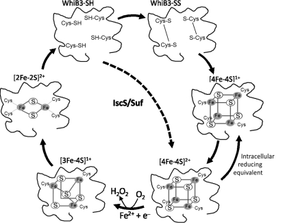
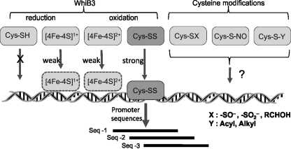
Recent advances: Recently, Mtb WhiB3, a member of the family of intracellular iron-sulfur (Fe-S) cluster proteins has emerged as a redox sensor and effector molecule controlling several aspects of Mtb virulence. WhiB3 was shown to contain a 4Fe-4S cluster that specifically reacts with important host gases (O(2) and NO), and exogenous and endogenous metabolic signals to maintain redox balance. Notably, the concept of reductive stress emerged from studies on WhiB3.

Critical issues: The detailed mechanism of how WhiB3 functions as an intracellular redox sensor is unknown. Sustaining Mtb redox balance is particularly important since the bacilli encounter a large number of redox stressors during infection, and because several antimycobacterial prodrugs are effective only upon bioreductive activation in the mycobacterial cytoplasm.

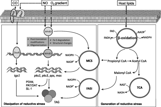
Future directions: How Mtb WhiB3 monitors its internal and external surroundings and modulates endogenous oxido-reductive pathways which in turn alter Mtb signal transduction, nucleic acid and protein synthesis, and enzymatic activation, is mostly unexplored. Modern expression, metabolomic and proteomic technologies should provide fresh insights into these yet unanswered questions.


Figures

FIG. 1.
FIG. 1.
Redox machinery of Mtb. During infection, Mtb encounters redox active compounds that have the capacity to alter or skew intracellular redox balance. Mtb has evolved several mechanisms to maintain redox homeostasis. Mtb utilizes host lipids as a source of carbon in vivo, which are metabolized via the (-oxidation pathway. This leads to the generation of high concentrations of NAD(P)H during the conversion of fatty acids to acetate and propionate. The accumulation of high levels of NAD(P)H causes reductive stress in Mtb (18, 40). Furthermore, host-generated free radicals such as reactive oxygen species (ROS) and reactive nitrogen species (RNS) are also capable of disturbing Mtb redox balance. However, Mtb counterbalances the stress generated by these radicals primarily via the abundant intracellular redox buffer, mycothiol (MSH). In addition, protein thiols and mycobacterial thioredoxins (Trx) are also involved in detoxifying the adverse effect of free radicals. Another yet uncharacterized redox active molecule, ergothioneine (Erg), is also expected to play a role in detoxification of free radicals and maintaining redox balance. Endogenous byproducts of mycobacterial respiration such as H2O2 and O2•− may react with mycobacterial lipids to generate lipid peroxides. In the presence of iron or copper, these ROS can generate the highly redox active OH. ROS or RNS such as peroxide, peroxynitrous acid (ONOOH), and peroxynitrite (ONOO-), and compounds such as vitamin C (in the presence of metal ions) may also lead to the production of intramycobacterial OH that can cause the oxidation of intracellular substrates. Host-generated gases also play a role in inducing mycobacterial redox changes. Mtb has well-defined sensor systems such as the DosS/T/R and WhiB3 signaling pathways, which specifically sense NO, CO, and changes in pO2. CO, NO, and O2 interact with the Dos signaling pathway, leading to the induction of the 47-member Dos regulon. It also leads to the production of the storage lipid TAG, which requires large quantities of NAD(P)H for synthesis. Similarly, WhiB3 also senses and responds to alterations in cytoplasmic NO and O2. Mrx; mycoredoxin, Mtr; mycothiol disulfide reductase.
FIG. 2.
FIG. 2.
Phylogenetic analysis of Mtb (A) WhiB3, (B) WhiB6, and (C) WhiB5. BLAST analyses were performed against the NCBI ‘Nr’ database. All the hits (except candidates from the same species) with e ≥1 e−10 were considered significant and further used in Clustal W alignment. A maximum likelihood method was used for tree construction, and boot strap values are indicated along the tree nodes. This analysis demonstrates that Mtb WhiB3 constitute a separate node (arrow) (A) from Streptomyces spp. and is closer related to Nocardia and Clostridium spp. A similar distribution pattern and tree architecture also hold true for Mtb WhiB1, WhiB2, WhiB4, and WhiB7 (not shown), but not for WhiB6 (B) and WhiB5 (C), which are restricted to mycobacteria. (To see this illustration in color the reader is referred to the web version of this article at www.liebertonline.com/ars).
FIG. 3.
FIG. 3.
Phylogenetic relationship among Mtb Wbl proteins and domain organization across species. (A) Mtb Wbl protein sequences were extracted from NCBI and aligned using Clustal W. In addition to four invariant Cys residues that are a characteristic signature of the Wbl family, the small G-X-W motif, typical of fatty acid binding proteins, is also present. Note that WhiB5 lacks a Cys residue. (B) Domain architecture of the Wbl family indicates that the “WhiB” domain is the most widely represented since it is present in ∼90% of Wbl members. All Streptomyces, mycobacterial phages, and mycobacterial WhiB1, WhiB2, WhiB4, WhiB5, and WhiB6 homologues share this architecture. (C) Phylogenetic tree was constructed using the maximum likelihood method and revealed that Mtb Wbl's are clustered in two nodes comprising of WhiB5 and WhiB6 in one node, and WhiB1, WhiB2, WhiB3 (red arrow), WhiB4, and WhiB7 in another. This indicates that WhiB5 and WhiB6 are functionally distinct from the other Mtb Wbl's. The numbers along the nodes denote bootstrap values, and the phylogenetic scale represents the number of differences between different sequences.
FIG. 4.
FIG. 4.
Relative transcription profiles of Mtb wbl genes. Graphic representation of the independent Mtb wbl transcription profiles extracted from published studies (9, 35, 45). WhiB3 and especially whiB6 appear to be induced under a wide range of conditions including starvation, hypoxia, and within macrophages. Expression of whiB4 was repressed under nutrient-starved conditions and upon infection of macrophages, whereas whiB4 expression was slightly induced during hypoxia. Notably, whiB1 and whiB2 appears to be strongly repressed under hypoxic conditions.
FIG. 5.
FIG. 5.
Diagrammatic representation of WhiB3 Fe-S cluster loss and assembly. In the presence of high concentrations of oxygen, WhiB3 [4Fe-4S]1+ cluster is oxidized to [4Fe-4S]2+ and is subsequently converted into a [3Fe-4S]1+ with the simultaneous generation of H2O2. The [3Fe-4S]1+ further yields [2Fe-2S]2+ intermediates, followed by a complete loss of the cluster wherein WhiB3 either exists in a reduced WhiB3-SH state, or oxidized WhiB3-SS state. Mtb IscS converts apo-WhiB3 to WhiB3 [4Fe-4S]2+. As suggested for Mtb WhiB1 (42), it is tempting to speculate that thioredoxin and mycothiol may be involved in stabilizing the WhiB3 [4Fe-4S] cluster.
FIG. 6.
FIG. 6.
Model depicting the mechanisms of Mtb WhiB3 DNA binding. WhiB3 DNA binding is influenced by the redox state of the Fe–S cluster (in the case of holo-WhiB3), or alternatively by the oxidation states of the Cys residues (in the case of apo-WhiB3). As previously demonstrated (40), oxidized apo-WhiB3 binds DNA strongly, whereas reduced apo-WhiB3 does not bind DNA at all, and holo-WhiB3 ([4Fe-4S]1+ or [4Fe-4S]2+) binds DNA weakly. It is reasonable to assume that the redox state of the mycobacterial cytoplasm would influence protonation and deprotonation of the WhiB3 Cys residues to affect DNA binding. Since Mtb experiences gradients of in vivo signals, an alteration in the concentration of effector molecules may lead to differential responses. It is therefore proposed that WhiB3 may sense and differentiate between effector molecules and their concentrations Fe–S cluster intermediates (3Fe-4S, 2Fe-2S), oxidation states of the Cys thiols, or via post-translational modifications of the Cys residues. These redox modifications likely dictate WhiB3 binding and modulation of its target genes. Since Cys residues within a protein can be modified to -SH, disulfide (-SS-), sulfenate (-SO), sulfinate (-SO2), or s-nitrosylated states, it is likely that these modifications could also modulate DNA-binding activity.
FIG. 7.
FIG. 7.
Diagram illustrating the role of Mtb WhiB3 in redox regulation. Mtb WhiB3 is a DNA-binding protein that senses changes in the environmental gases such as NO and gradients of O2. WhiB3 also senses or responds to fluctuating levels of reducing equivalents (NADH) that are generated by the TCA cycle (respiration), and NADPH that accumulates during (β-oxidation of host fatty acids. If not properly balanced, the bacillus will experience reductive stress (18). As a result, WhiB3 channels these excess reducing equivalents into virulence lipid anabolic pathways, which requires NADPH as cofactor. Thus, virulence lipid anabolism (e.g., SL-1, PDIM, PAT, DAT, and trehalose dimycolate) functions as an electron sink or reserve storage material, which is available for future use. This is also, in part, a mechanism to maintain intracellular redox balance. WhiB3 also overlaps with the DosR/S/T signaling pathway since both regulates TAG synthesis.

Similar articles

Cited by

References

    1. Agarwal N. Raghunand TR. Bishai WR. Regulation of the expression of whiB1 in Mycobacterium tuberculosis: Role of cAMP receptor protein. Microbiology. 2006;152:2749–2756. - PubMed
    1. Alam MS. Garg SK. Agrawal P. Molecular function of WhiB4/Rv3681c of Mycobacterium tuberculosis H37Rv: A [4Fe-4S] cluster co-ordinating protein disulphide reductase. Mol Microbiol. 2007;63:1414–1431. - PubMed
    1. Alam MS. Garg SK. Agrawal P. Studies on structural and functional divergence among seven WhiB proteins of Mycobacterium tuberculosis H37Rv. FEBS J. 2009;276:76–93. - PubMed
    1. Aravind L. Landsman D. AT-hook motifs identified in a wide variety of DNA-binding proteins. Nucleic Acids Res. 1998;26:4413. - PMC - PubMed
    1. Banaiee N. Jacobs WR., Jr. Ernst JD. Regulation of Mycobacterium tuberculosis whiB3 in the mouse lung and macrophages. Infect Immun. 2006;74:6449–6457. - PMC - PubMed

Publication types

MeSH terms

Substances

LinkOut - more resources