Review
. 2012 Mar 20;45(3):444-53.
doi: 10.1021/ar200166p. Epub 2011 Oct 19.

Structure and mechanisms of Escherichia coli aspartate transcarbamoylase

Affiliations
Review

Structure and mechanisms of Escherichia coli aspartate transcarbamoylase

William N Lipscomb et al. Acc Chem Res. .

Abstract

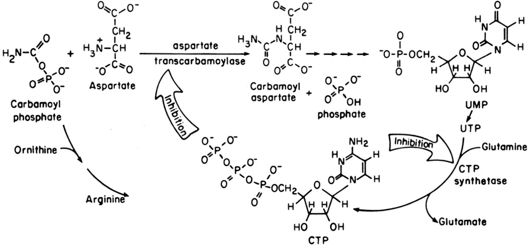
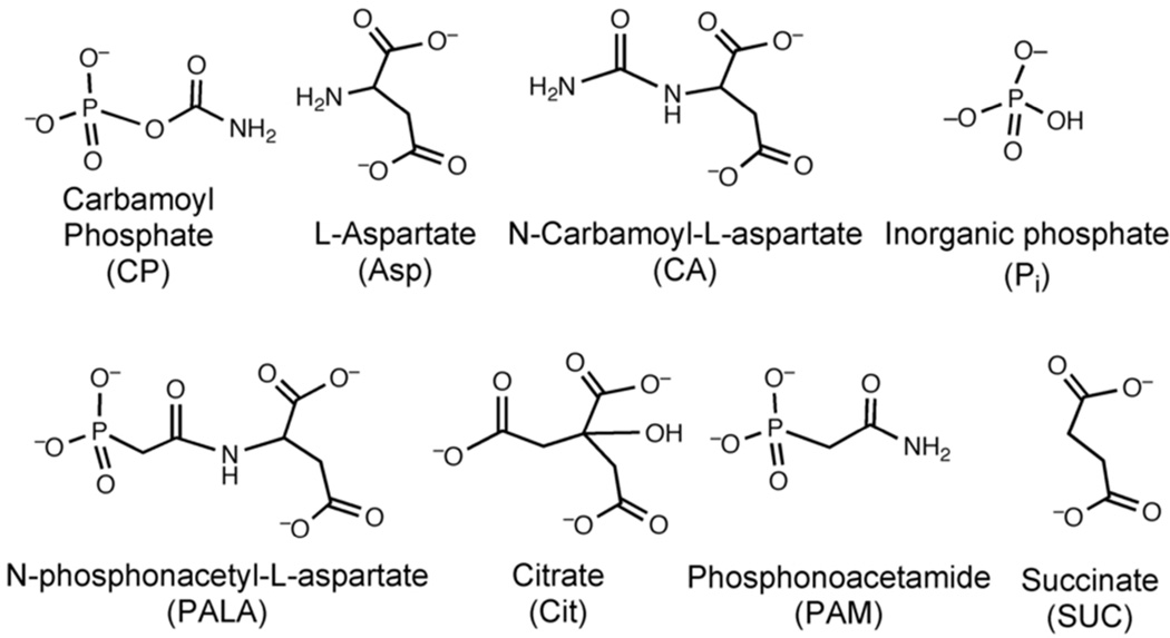
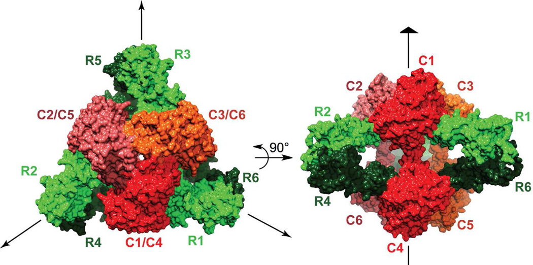
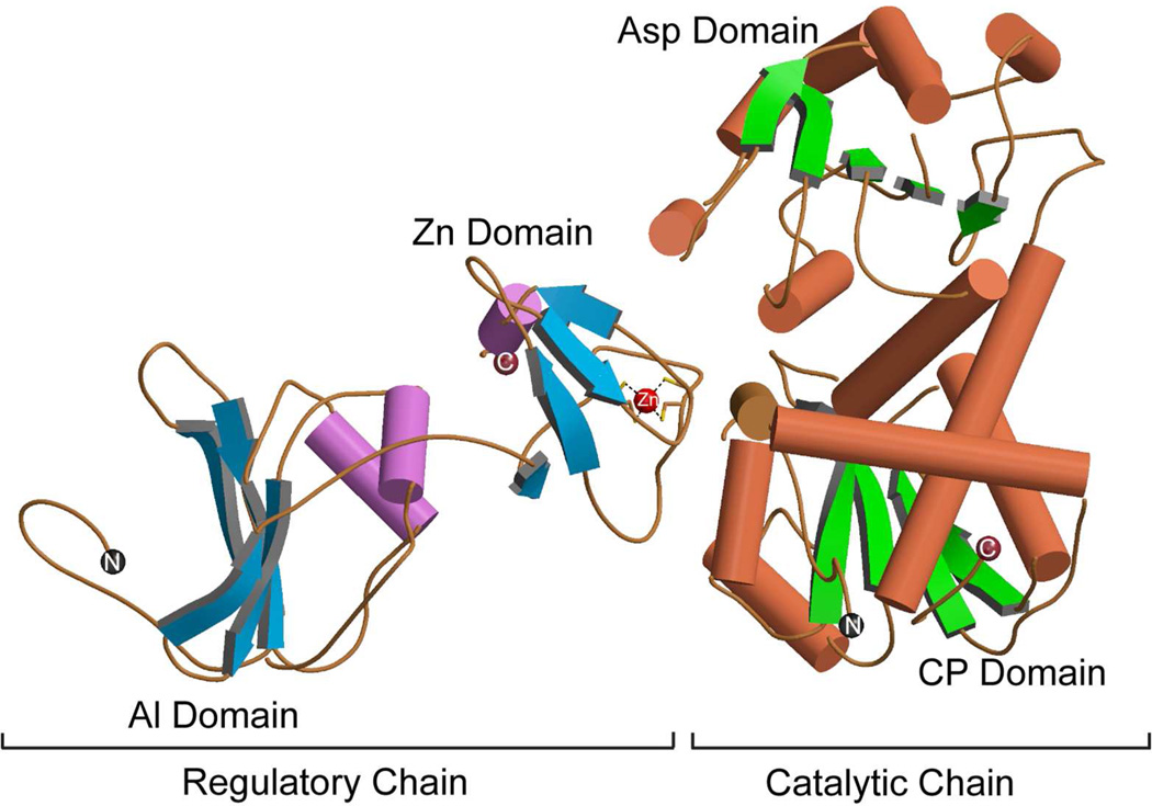
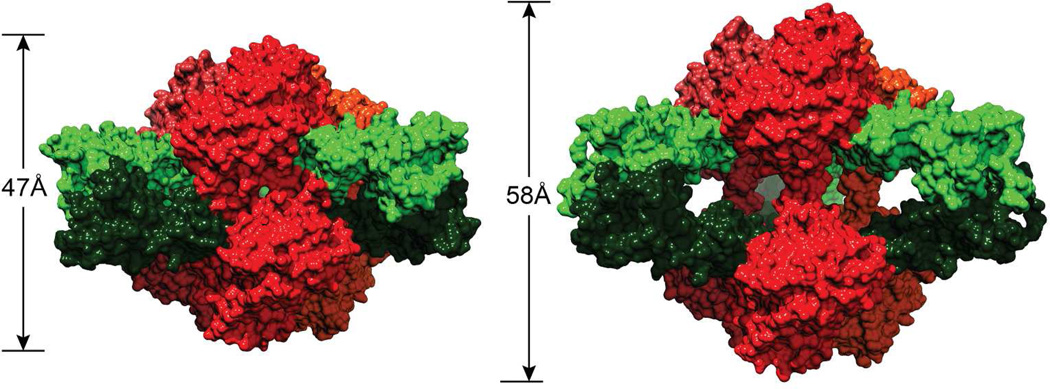
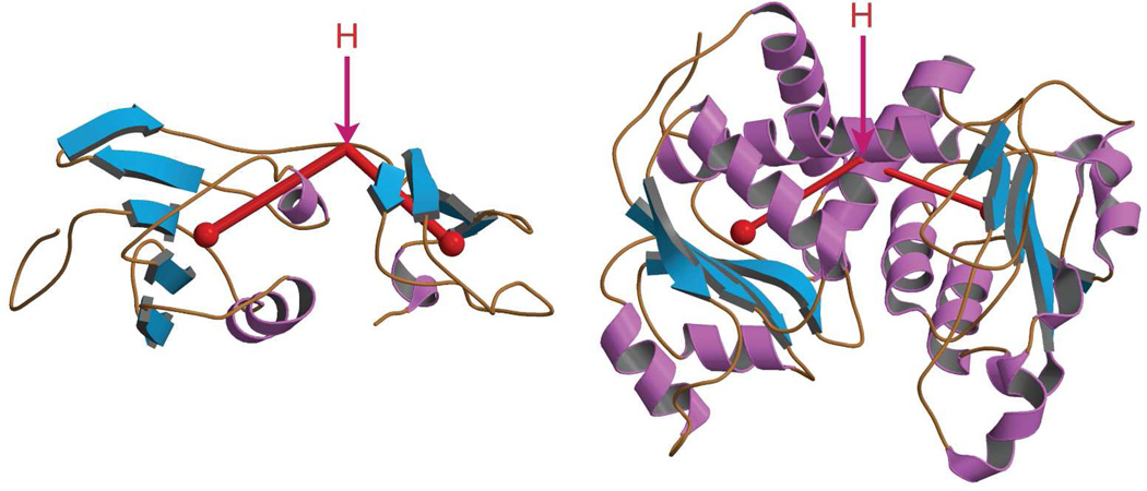
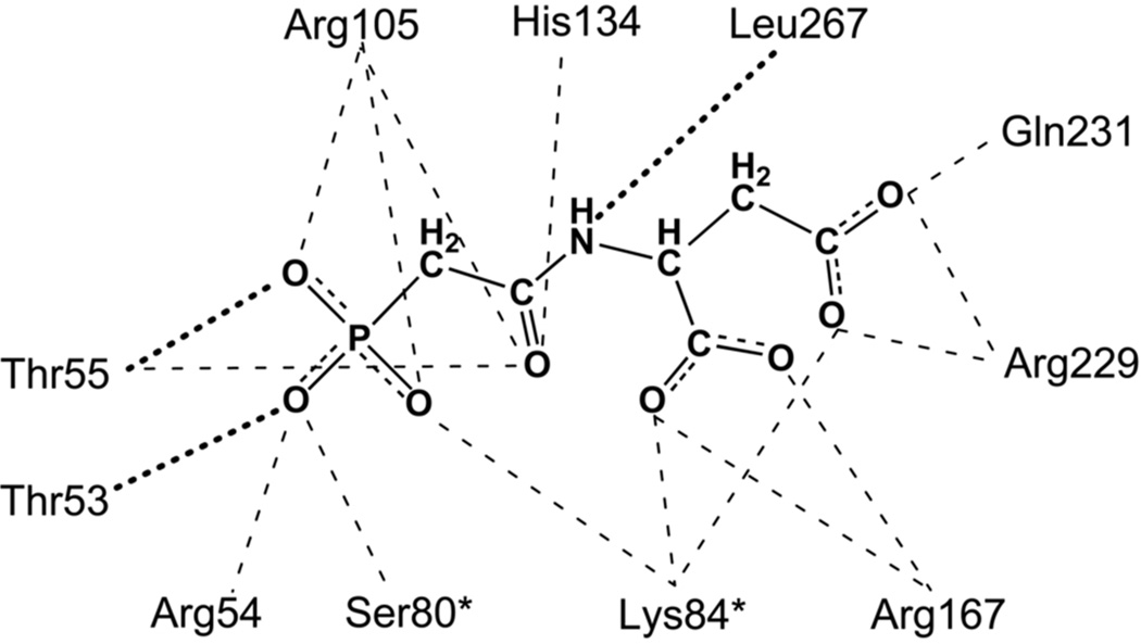
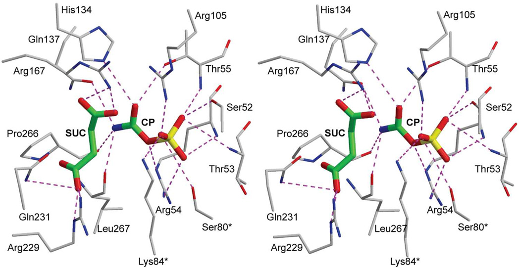
Enzymes catalyze a particular reaction in cells, but only a few control the rate of this reaction and the metabolic pathway that follows. One specific mechanism for such enzymatic control of a metabolic pathway involves molecular feedback, whereby a metabolite further down the pathway acts at a unique site on the control enzyme to alter its activity allosterically. This regulation may be positive or negative (or both), depending upon the particular system. Another method of enzymatic control involves the cooperative binding of the substrate, which allows a large change in enzyme activity to emanate from only a small change in substrate concentration. Allosteric regulation and homotropic cooperativity are often known to involve significant conformational changes in the structure of the protein. Escherichia coli aspartate transcarbamoylase (ATCase) is the textbook example of an enzyme that regulates a metabolic pathway, namely, pyrimidine nucleotide biosynthesis, by feedback control and by the cooperative binding of the substrate, L-aspartate. The catalytic and regulatory mechanisms of this enzyme have been extensively studied. A series of X-ray crystal structures of the enzyme in the presence and absence of substrates, products, and analogues have provided details, at the molecular level, of the conformational changes that the enzyme undergoes as it shifts between its low-activity, low-affinity form (T state) to its high-activity, high-affinity form (R state). These structural data provide insights into not only how this enzyme catalyzes the reaction between l-aspartate and carbamoyl phosphate to form N-carbamoyl-L-aspartate and inorganic phosphate, but also how the allosteric effectors modulate this activity. In this Account, we summarize studies on the structure of the enzyme and describe how these structural data provide insights into the catalytic and regulatory mechanisms of the enzyme. The ATCase-catalyzed reaction is regulated by nucleotide binding some 60 Å from the active site, inducing structural alterations that modulate catalytic activity. The delineation of the structure and function in this particular model system will help in understanding the molecular basis of cooperativity and allosteric regulation in other systems as well.


Figures

FIGURE 1
FIGURE 1
Aspartate transcarbamoylase (ATCase) catalyzes the committed step, the condensation of carbamoyl phosphate and aspartate to form carbamoyl aspartate and inorganic phosphate, in pyrimidine nucleotide biosynthesis for E. coli. Carbamoyl aspartate continues through the pathway leading to the formation of the nitrogenous base in pyrimidine nucleotides. ATCase is feedback inhibited by CTP and the combination of CTP plus UTP. This figure has been reproduced here with permission. Note to editor: I have obtained rights to reuse this figure via Rightslink.
FIGURE 2
FIGURE 2
The structures of carbamoyl phosphate (CP), L-aspartate (Asp), N-carbamoyl-L-aspartate (CA), phosphate (Pi), the bisubstrate analogue N-phosphonacetyl- L-aspartate (PALA), the N-carbamoyl-L-aspartate analogue citrate (Cit), the CP analogue phosphonoacetamide (PAM), and the Asp analogue succinate (SUC).
FIGURE 3
FIGURE 3
Schematic representation of the quaternary structure of ATCase. Catalytic chains C1-C2-C3 and C4-C5-C6 correspond to the two catalytic trimers while the regulatory chains R1–R6, R2–R4 and R3–R5 correspond to the three regulatory dimers. This figure was drawn with Chimera.
FIGURE 4
FIGURE 4
The secondary structural elements of one catalytic and one regulatory chain of E. coli ATCase in the T-state. The N- and C-termini of the catalytic and regulatory chains are indicated, as well as the Zn atom in the regulatory chain that is coordinated tetrahedrally to four cysteine residues. Each catalytic chain is composed of an aspartate (Asp) and a carbamoyl phosphate (CP) domain. Each regulatory chain is composed of an allosteric (Al) and a zinc (Zn) domain. This figure was drawn with MOLSCRIPT using data from PDB entry 1ZA1.
FIGURE 5
FIGURE 5
The ATCase holoenzyme in the T (left) and R (right) structures. The catalytic chains are shown in shades of red and the regulatory chains are shown in shades of green. The length of the molecule along the three-fold axis is shorter in the T form than in the R form. This figure was drawn with Chimera.
FIGURE 6
FIGURE 6
Secondary structure of a regulatory chain (left) and a catalytic chain (right) of ATCase. The angle between the domains is measured from the center of mass of each domain (red dot) and a hinge point (H).
FIGURE 7
FIGURE 7
The interactions between PALA and ATCase in the R-state. Dotted lines correspond to interactions with the backbone of the protein. An asterisk after the residue number indicates that it is donated into the active site from the adjacent chain.
FIGURE 8
FIGURE 8
Stereoview of succinate (SUC) and CP bound in the active site of ATCase in the R state. An asterisk after the residue number indicates that it is donated into the active site from the adjacent chain. This figure was drawn with MOLSCRIPT.
FIGURE 9
FIGURE 9
Stereoview of the proposed tetrahedral intermediate (TET-I) bound in the active site of ATCase in the R state. An asterisk after the residue number indicates that it is donated into the active site from the adjacent chain. This figure was drawn with MOLSCRIPT.
FIGURE 10
FIGURE 10
Stereoview of citrate (CIT) and phosphate (Pi) bound in the active site of ATCase in the R state. An asterisk after the residue number indicates that it is donated into the active site from the adjacent chain. This figure was drawn with MOLSCRIPT.
FIGURE 11
FIGURE 11
Stereoview comparison of the active site of E. coli ATCase in the presence of CP (green carbons, thick) and in the absence of CP (black carbons, thin). The 80's loop from the adjacent chain (residue numbers with asterisks] in the presence of CP (gold carbons, thick) and in the absence of CP (gray carbons, thin) is represented as a coil with Ser80* and Lys84* shown. This figure was drawn with MOLSCRIPT.
FIGURE 12
FIGURE 12
(A) Electrostatic potentials mapped onto the surface of ATCase in the absence (A) and the presence (B) of CP. The positions of Ser80*, Lys84* and Arg234 and CP are overlaid onto the electrostatic map (white carbons, sticks). This figure has been reproduced here with permission. Note to editor [no copyright transfer needed for author to reuse figure PNAS, 102, 8881–8886 (2005)]
FIGURE 13
FIGURE 13
Surface of ATCase near the active site detailing the 80's loop (green) and the 240's loop (red) during catalysis. (A) Conformation of the enzyme before substrates are bound; the approximate position of the active site is indicated by the magenta arrow. (B) Conformation of the enzyme after the binding of CP, which is shown in the active site as a CPK model (white carbon). The 80's loop (green) rearranges to help create the binding site for aspartate. (C) Upon the binding of aspartate the 240's loop (red) undergoes a dramatic conformational change, forcing the substrates toward each other. Solvent is not accessible to the active site in this conformation.
FIGURE 14
FIGURE 14
A comparison of the interactions between CTP (top) and ATP (bottom) and the allosteric site on the regulatory chains of E. coli ATCase. This figure is based on the ATCase•CTP and ATCase•ATP structures in the T state.

Similar articles

Cited by

References

    1. Yates RA, Pardee AB. Control of pyrimidine biosynthesis in Escherichia coli. J. Biol. Chem. 1956;221:757–770. - PubMed
    1. Umbarger HE. Evidence for a Negative-Feedback Mechanism in the Biosynthesis of Isoleucine. Science. 1956;123:848–848. - PubMed
    1. Shepherdson M, Pardee AB. Production and Crystallization of Aspartate Transcarbamylase. J. Biol. Chem. 1960;235:3233–3237.
    1. Jones ME, Spector L, Lipmann F. Carbamyl phosphate. The carbamyl donor in enzymatic citrulline synthesis. J. Am. Chem. Soc. 1955;77:819–820.
    1. Lowenstein JM, Cohen PP. Studies on the Biosynthesis of carbamylaspartic acid. J. Biol. Chem. 1956;235:57–78. - PubMed

Publication types

MeSH terms

Substances